Clear Sky Science · en
Structural basis for the differential recognition of integrin αvβ3 by rhodostomin and trimucrin
Venom Proteins Turned Into Precision Medicine Tools
Snake venom may sound like pure poison, but some of its molecules are remarkably good at blocking disease-related proteins in our bodies. This study explores how two such venom-derived molecules latch onto a key cell-surface receptor linked to cancer and blood vessel growth, revealing atomic-level details that could guide the design of safer, more precise drugs.
A Cell Grip That Cancer Learns to Exploit
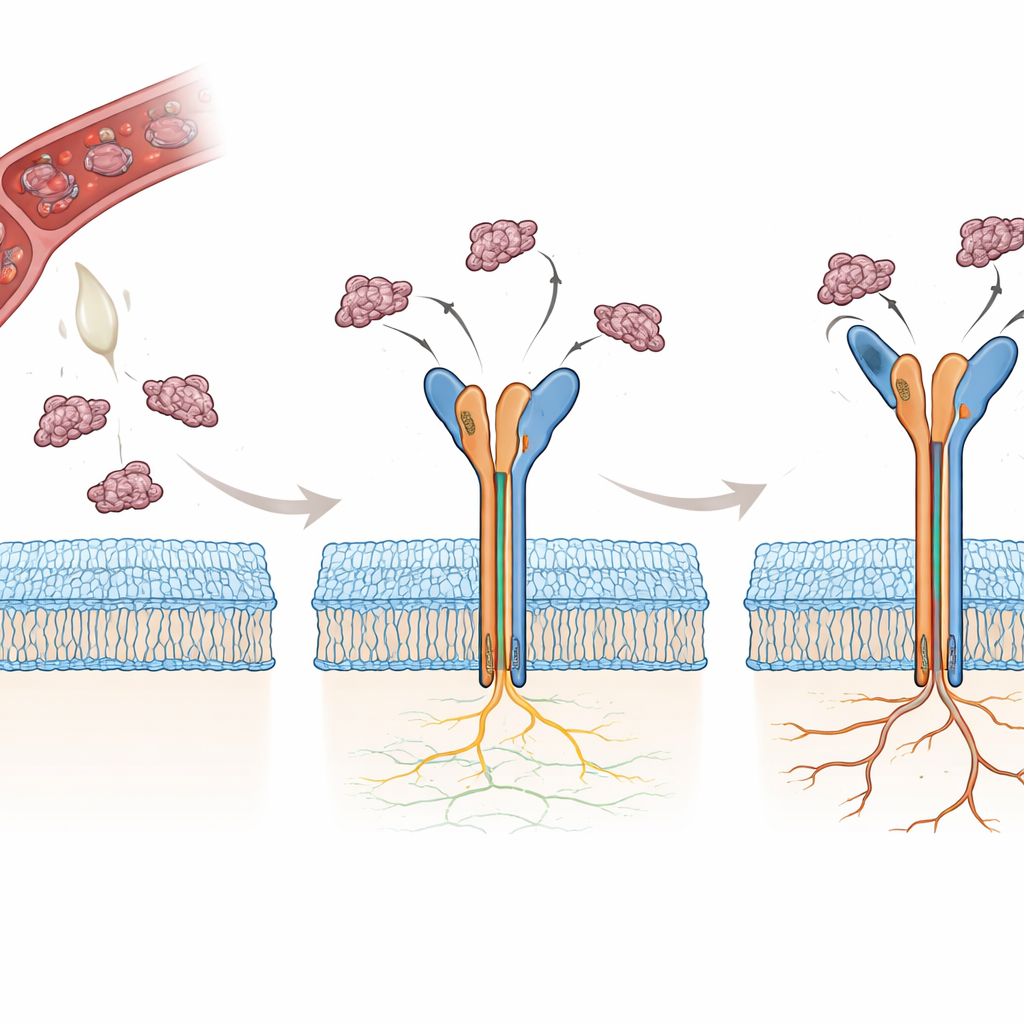
Our cells use protein "grips" called integrins to attach to their surroundings and to sense mechanical and chemical cues. One particular integrin, called αvβ3, helps cells respond to their environment and plays a major role in the growth of new blood vessels, tumor expansion, spread of cancer cells, and drug resistance. Because of this, αvβ3 has been a long-standing target for anti-cancer and anti-angiogenic drugs. Yet decades of work have produced no approved αvβ3 blockers for human use, in part because it has been difficult to understand exactly how different molecules bind to and control this receptor.
Why Snake Venom Molecules Are So Potent
Many snakes produce small proteins called disintegrins that naturally block integrins, preventing blood from clotting and interfering with cell adhesion. Two such disintegrins, rhodostomin (Rho) and trimucrin (Tmu), both contain a short sequence of three amino acids—arginine-glycine-aspartate, or RGD—that is recognized by αvβ3. Surprisingly, Tmu is about three to four times better than Rho at shutting down this integrin. The two molecules share most of their sequence but differ in three key regions: a short linker, the flexible RGD-containing loop, and the tail end of the protein. The authors set out to determine, atom by atom, how these small differences change the way each venom protein grips αvβ3 and why that makes Tmu a more powerful inhibitor.
Capturing the Structures in High Detail
To answer these questions, the researchers first solved the crystal structure of Tmu alone at very high resolution, confirming that it forms a compact, rigid framework stabilized by six disulfide bonds. They then used cryogenic electron microscopy to determine three-dimensional structures of αvβ3 bound to either Rho or Tmu, reaching resolutions under three angstroms for the head of the receptor where binding occurs. In both cases, the disintegrin sits in a crevice between two domains of the integrin head, inserting its RGD loop into a metal-containing site. The backbone shape of Rho and Tmu in this region is almost identical, showing that both proteins arrive pre-formed in a binding-ready shape, rather than having to undergo large rearrangements when engaging the receptor.
Three Contact Zones and a Stronger Clamp
Closer inspection revealed that binding is not governed by the RGD triplet alone. Both disintegrins use three cooperative contact zones: the RGD loop itself; a nearby linker region; and the C-terminal tail. In Tmu, a cluster of positively charged amino acids in the linker makes additional electrostatic contacts with the negatively charged surface of the β3 subunit, helping it clamp more tightly onto αvβ3. Rho, by contrast, relies more on its tail region for extra contacts. When the team swapped specific sequence segments between the two venom proteins, they could dial the blocking power up or down. In particular, transferring Tmu’s charged linker into Rho boosted Rho’s ability to inhibit αvβ3 by roughly tenfold, confirming that this extra binding patch is a major contributor to potency.
How Binding Flips the Receptor On—and How a Single Atom Matters
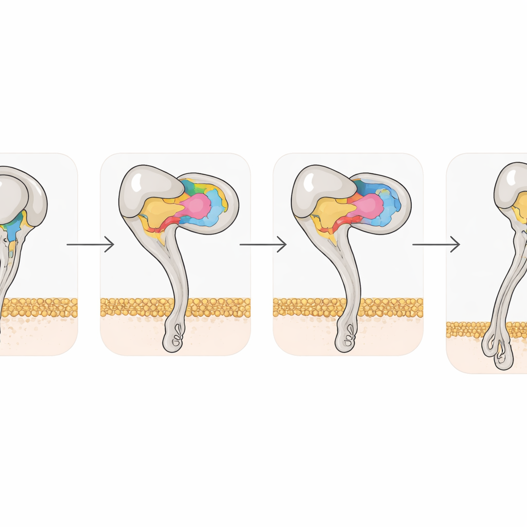
The structural snapshots also captured how disintegrin binding reshapes the integrin itself. Free αvβ3 prefers a bent, closed posture; once Rho or Tmu binds, the β3 half of the receptor undergoes coordinated shifts in two helices and metal-binding sites, driving the receptor into a fully extended, open state associated with high activity. Yet a single amino acid, tyrosine 110 in β3, helps hold the bent form together by bridging two domains. When the researchers mutated this residue to alanine, the receptor spontaneously favored the extended state and bound more strongly to its natural partner, vitronectin. This highlights how subtle atomic changes can tune integrin activation and suggests that engineered variants could be useful tools for screening new drugs.
From Venom Clues to Future Cancer Drugs
Taken together, the work shows that medium-sized venom disintegrins provide a highly efficient scaffold for targeting αvβ3, using three coordinated contact regions and a rigid binding loop to outperform simple RGD-based molecules. By mapping exactly how Tmu and Rho engage the receptor—and how small sequence changes alter this engagement—the study offers a structural blueprint for designing next-generation integrin blockers that are more selective for αvβ3 and less likely to cause bleeding or other side effects. In essence, by learning how snakes disable our cell adhesion machinery with exquisite precision, scientists gain new ideas for turning these natural weapons into targeted therapies against cancer and inflammatory disease.
Citation: Wang, YT., Chang, YT., Huang, CH. et al. Structural basis for the differential recognition of integrin αvβ3 by rhodostomin and trimucrin. Commun Biol 9, 583 (2026). https://doi.org/10.1038/s42003-026-10139-6
Keywords: integrin alpha v beta 3, snake venom disintegrins, rhodostomin trimucrin, cancer and angiogenesis, cryo electron microscopy