Clear Sky Science · en
Real-time quantification during indocyanine green fluorescent-guided surgery in canine soft tissue sarcomas and mast cell tumors
Why glowing tumors matter for dogs
For many dog owners, cancer surgery is a race against time and uncertainty. Surgeons must remove all cancer while sparing as much healthy tissue as possible, yet tumor borders can be hard to see with the naked eye. This study tests a light-based method that makes tumors glow during surgery, helping veterinarians see where cancer ends and healthy tissue begins in real time.
A dye that makes cancer stand out
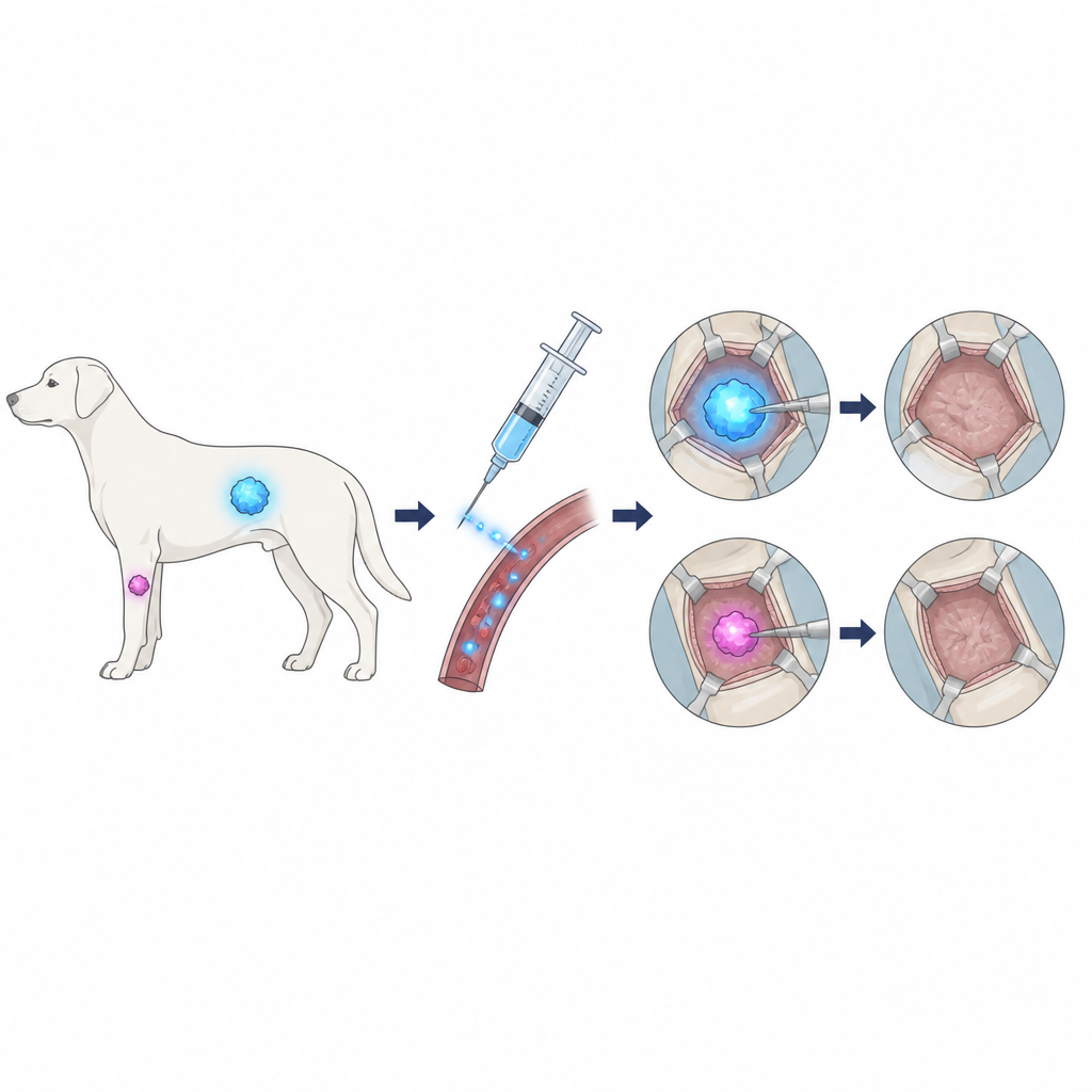
The research team focused on two common cancers in dogs: soft tissue sarcomas, which grow in muscles and connective tissue, and mast cell tumors, which often arise in the skin and just beneath it. They used a medical dye called indocyanine green, already employed in human hospitals, that travels through the bloodstream and tends to collect more in tumor tissue than in normal tissue. When illuminated with near-infrared light, the dye glows, revealing areas with higher cancer content. The scientists wanted to know whether this glow could reliably guide surgery in real dogs with naturally occurring tumors, not just in experimental models.
How the glowing-guided surgery was done
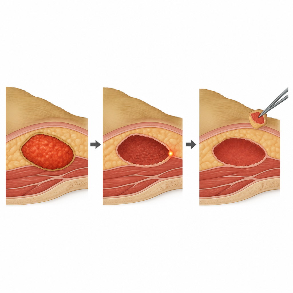
Twenty-six pet dogs with a total of 28 tumors were enrolled, all scheduled for surgery with the aim of cure. The dogs received a low dose of the dye through a vein 24 hours before the operation. During surgery, the team used a handheld camera system that could switch between normal white-light view and a special mode that detects the dye’s glow. They continually measured how bright the tumor was compared with nearby apparently normal tissue before cutting, while removing the tumor, and again after the tumor was out, examining the wound bed where it had been. If certain areas glowed more strongly than the surrounding tissue, surgeons were prompted to remove a small extra rim of tissue from that spot when it was safe to do so.
What the surgeons and pathologists found
The dye-guided method worked in all 14 soft tissue sarcomas and in 11 of 14 mast cell tumors; three mast cell tumors simply did not glow more than the background, so the technique could not help in those cases. Overall, tumors were much brighter than their surroundings, especially the sarcomas. Surgeons extended their planned cuts in about two thirds of sarcomas and over half of mast cell tumors because of the fluorescence signal, usually by taking an extra half to nearly one centimeter of tissue at one edge. After the main tumor was removed, roughly half of the wound beds still showed glowing spots; in most of these, extra tissue was taken for safety, provided this would not damage important structures like nerves or blood vessels.
How well did glow match hidden cancer?
To test whether glowing areas truly meant lingering cancer, all removed tissue and wound samples were examined under the microscope. In sarcomas, the glow in the wound bed matched infiltrated, or cancer-positive, edges fairly well. When the wound glow suggested remaining sarcoma, this was correct in many cases, and when there was no glow, there was a high chance the edges were truly clean. In contrast, mast cell tumors showed a weaker link between glow and microscopic spread. The dye sometimes lit up tissues that turned out to be non-cancerous, and it sometimes missed small clusters of remaining mast cells. The authors suggest that the biology of mast cell tumors, including local inflammation and leakage of blood vessels, may cause the dye to gather outside the true tumor zone, blurring the signal.
What this means for dogs and their vets
This pilot study shows that making tumors glow during surgery is safe and technically workable in everyday veterinary practice, especially for soft tissue sarcomas. For these tumors, the method can help surgeons decide, on the spot, where to take a little more tissue and where it is likely safe to stop cutting. For mast cell tumors the tool is less reliable on its own, and surgeons must be more cautious in how they interpret the glow. Overall, the work points toward a future where real-time visual cues in the operating room can improve the thoroughness of cancer surgery in dogs, while larger studies refine the technique and tailor it to each tumor type.
Citation: Gariboldi, E.M., Ubiali, A., Luconi, E. et al. Real-time quantification during indocyanine green fluorescent-guided surgery in canine soft tissue sarcomas and mast cell tumors. Sci Rep 16, 16178 (2026). https://doi.org/10.1038/s41598-026-47495-6
Keywords: canine cancer, fluorescence-guided surgery, soft tissue sarcoma, mast cell tumor, indocyanine green