Clear Sky Science · en
Role of ethnicity in the determination of the transverse sigmoid sinus Junction (TSSJ): A prospective hospital-based radiological study in adult Sabah, East Malaysia
Why the Shape of Our Skulls Matters in Brain Surgery
When surgeons operate near the base of the brain, just a few millimetres can mean the difference between a safe procedure and serious bleeding. This study from Sabah, East Malaysia, asks a deceptively simple question: does a person’s ethnic background—and the way it subtly shapes the skull—change where a critical vein junction lies beneath the bone? By answering this, the researchers hope to make delicate brain operations safer, especially in hospitals that cannot rely on advanced navigation machines.
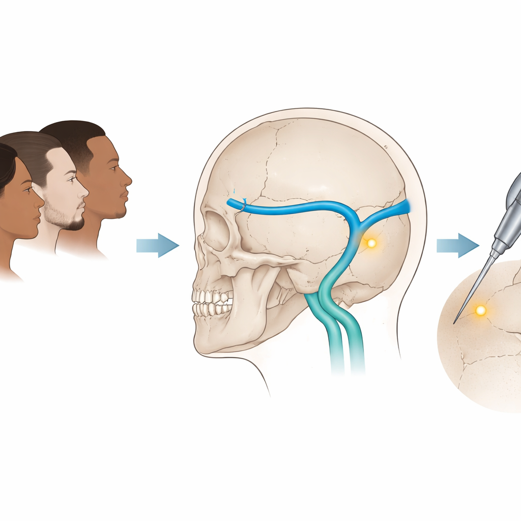
A Hidden Junction Deep Behind the Ear
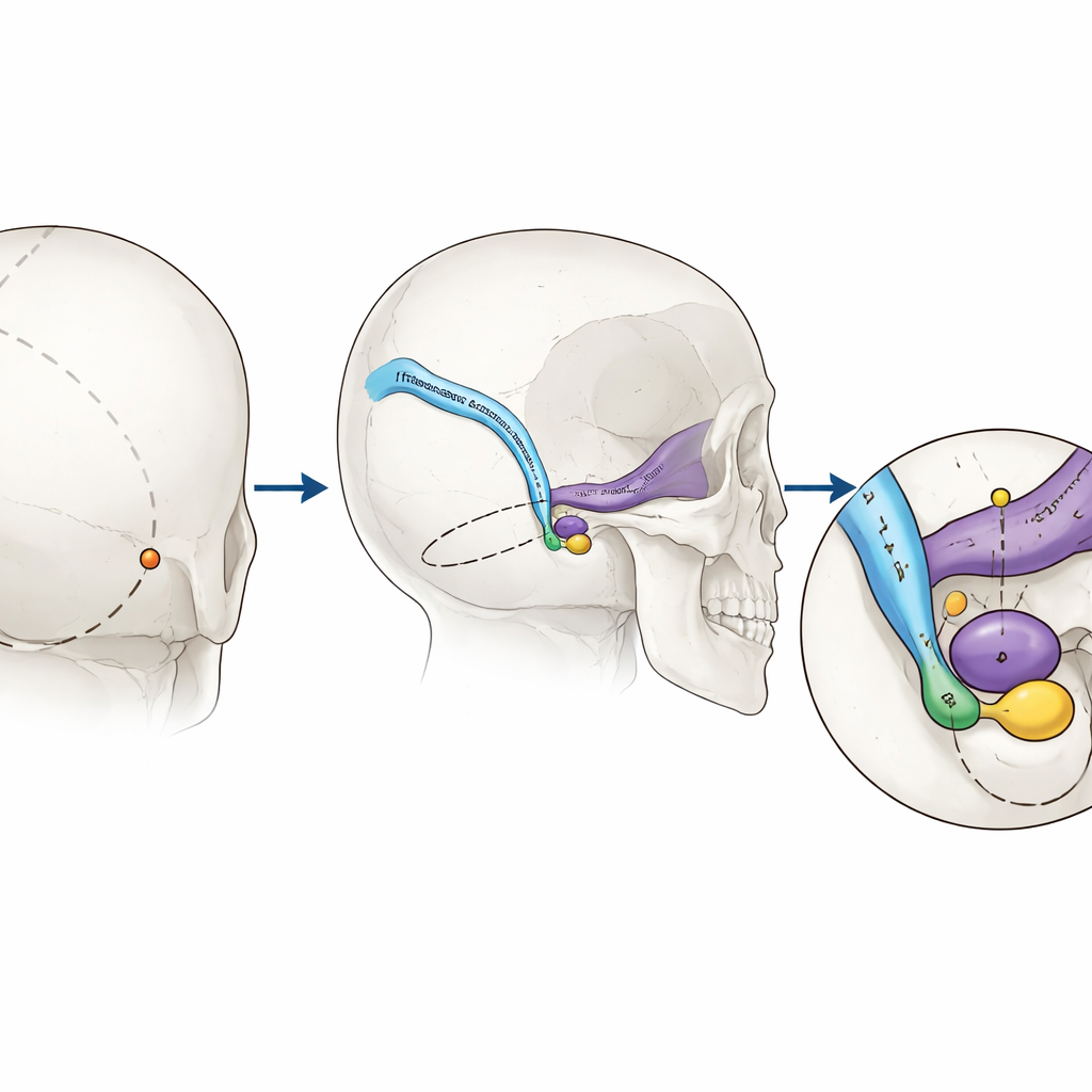
Behind each ear, large veins drain blood from the brain as they curve from a horizontal channel (the transverse sinus) into a vertical one (the sigmoid sinus). The bend where these two meet—the transverse–sigmoid sinus junction—is a key landmark for operations in the cerebello-pontine angle, a crowded region near the brainstem. Surgeons often make a small opening, or burr hole, in the skull close to this junction. If they place it too far forward or too low, they risk cutting into the vein. Traditionally, they rely on surface landmarks on the skull, such as a point where several seams in the bone meet, called the asterion. But past work has shown these landmarks are not in the same place in everyone, raising concerns about how much surgeons can trust them.
Scanning the Skulls of a Diverse Population
Sabah is home to more than 50 ethnic communities with distinct ancestry. To see how this diversity might affect surgical landmarks, the team analysed high‑resolution CT scans from 180 adults aged 22 to 80 who had already undergone imaging for clinical reasons. Using specialised 3D software, they reconstructed each person’s skull and venous system. They then located a “key point” on the skull surface that lies directly over the junction of the two sinuses, defined by precise geometric lines drawn along the veins inside the bone. From this key point, they measured distances to the asterion and to the internal auditory canal, a short tunnel in the bone that leads nerves and vessels into the inner ear. They also calculated a cranial index—a simple ratio describing whether a skull is relatively long and narrow or short and broad.
Differences Between Left and Right, Men and Women
The researchers found that the key point was not perfectly symmetrical between the two sides of the head. On average, the distance from the asterion to the key point was very slightly greater on the left than on the right, although the difference was only fractions of a millimetre and probably of limited practical impact. Sex differences, on the other hand, were much more striking. Men tended to have larger skull dimensions overall and consistently greater distances from the asterion to the key point, from the key point to the internal auditory canal, and in the vertical separation between these structures. In the tight confines of posterior fossa surgery, where surgeons work within a few millimetres of major veins, a shift of three to four millimetres in these relationships can meaningfully alter both access and risk.
How Ethnic Background and Skull Shape Come Into Play
When the team compared ethnic groups—Kadazan, Malay, Chinese, and a combined “Others” group—they did see statistically significant differences, but these were modest in size for the raw distances along the skull. Chinese participants, for instance, had a somewhat greater asterion‑to‑key point distance than those grouped as “Others.” The most pronounced difference across groups lay in the cranial index: people in the “Others” category, which included several indigenous groups such as Bajau and Murut, tended to have longer, narrower skulls. These overall shape differences were linked to where the key point and venous junction sat, suggesting that how broad or elongated a skull is may matter more for surgical planning than a simple label of ethnic group.
Why These Findings Matter for Patient Safety
In many well‑resourced hospitals, surgeons can rely on real‑time navigation systems that map instruments to a patient’s scans during surgery. But such technology is not always available, and even when it is, it can lose accuracy as the brain shifts during an operation. This study shows that using “one‑size‑fits‑all” surface landmarks on the skull can be risky in a multi‑ethnic population like Sabah’s, where skull shapes, and thus the positions of hidden veins, vary with sex and ancestry. The authors argue that careful, individual measurements from pre‑operative scans—rather than assumptions based on standard anatomy—should guide where surgeons place their openings. In other words, tailoring neurosurgical planning to each person’s unique cranial shape can help avoid damage to vital veins and make complex brain procedures safer.
Citation: Naesarajoo, J.J.J., Abdullah, J.Y., Avoi, R. et al. Role of ethnicity in the determination of the transverse sigmoid sinus Junction (TSSJ): A prospective hospital-based radiological study in adult Sabah, East Malaysia. Sci Rep 16, 14458 (2026). https://doi.org/10.1038/s41598-026-44484-7
Keywords: neurosurgery, skull anatomy, ethnic variation, brain venous sinuses, medical imaging