Clear Sky Science · en
The EHMT2-MBLAC2 axis suppresses ribosomal DNA transcription in response to nucleolar DNA damage
Why the cell’s protein factories need protection
Inside each of our cells lies a bustling workshop called the nucleolus, where the molecular machines that make proteins—ribosomes—are built. Because this factory runs almost nonstop, its DNA template is under constant strain and can break. The study in this article explores how cells temporarily quiet this factory to repair damage, and how failure in this safety system may help drive colorectal cancer.
A vulnerable stretch of DNA
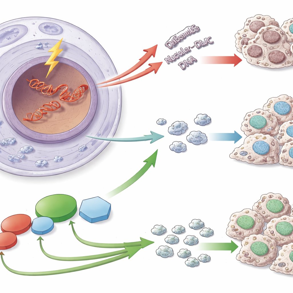
Ribosomes are assembled using instructions encoded in special DNA segments known as ribosomal DNA (rDNA). These sequences are repeated many times and are copied at very high speed, which makes them especially prone to dangerous double-strand breaks. When such breaks occur in rDNA, cells do more than just patch the DNA: they also reorganize the nucleolus and briefly shut down the copying of rDNA into RNA. This pause prevents repair machines from colliding with the transcription machinery and helps keep the number and structure of rDNA repeats stable—an important safeguard, because rDNA instability has been linked to aging and many diseases, including cancer.
A new safety switch in the nucleolus
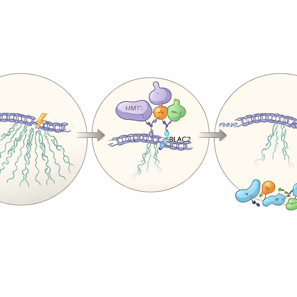
The researchers identify a protein called EHMT2, better known for modifying chromatin elsewhere in the genome, as a crucial player in this nucleolar safety response. Using a system in human cells that cuts rDNA at defined sites, they show that EHMT2 rushes to the edge of the nucleolus where damaged rDNA is gathered into structures called caps. There, EHMT2 is required to shut down ribosomal RNA production after damage. When EHMT2 is missing or its activity is chemically blocked, cells continue to transcribe rDNA despite the breaks. The team traces this effect to EHMT2’s ability to add a specific chemical mark to histone proteins at damaged rDNA, helping to create a more repressed chromatin state.
Linking transcription pause to DNA repair
Quieting rDNA is only half the story; successful repair must follow. The authors find that cells lacking EHMT2 struggle to repair rDNA breaks: DNA fragments persist longer, small extra nuclei (a sign of genomic instability) appear more frequently, and the number of rDNA copies declines over time. Importantly, one major repair pathway—non-homologous end joining, which rapidly reseals DNA ends—fails to be properly recruited to rDNA caps without EHMT2, even though other repair routes remain largely intact. As a result, EHMT2-deficient cells become hypersensitive to nucleolar damage and lose their ability to form healthy colonies after rDNA is cut.
A hidden partner: MBLAC2
To understand how EHMT2 exerts its influence, the team performs large-scale protein measurements in cells with and without EHMT2. Among the proteins that drop when EHMT2 is absent, one stands out: MBLAC2, a little-studied enzyme normally enriched in the nucleolus. When MBLAC2 is reduced with targeted RNA tools, cells again fail to silence rDNA after damage and become less able to survive it. EHMT2 physically binds to MBLAC2 and stabilizes it at the protein level; without EHMT2, MBLAC2 is degraded more quickly. Removing both proteins does not worsen the effect beyond removing one, suggesting they act in the same pathway—a newly described EHMT2–MBLAC2 axis that coordinates the transcription pause with effective repair.
From damaged DNA to colorectal cancer
Because rDNA instability is a hallmark of many tumors, the authors ask whether this axis plays a role in colorectal cancer, a disease that strongly depends on elevated ribosome production. In mouse models and human tumor samples, both EHMT2 and MBLAC2 are consistently more abundant in colorectal tumors than in normal colon tissue, in parallel with markers of boosted ribosome formation. Suppressing EHMT2 in colorectal cancer cells slows the growth of tumors in mice, reduces ribosome-related markers, and lowers MBLAC2 levels. Patient data from The Cancer Genome Atlas reveal that higher expression of both genes associates with more advanced disease and poorer survival after radiotherapy. Together, these observations suggest that cancer cells may hijack the EHMT2–MBLAC2 pathway to better tolerate rDNA damage and maintain the ribosome output needed for uncontrolled growth.
What this means for health and therapy
To a lay reader, the key message is that our cells have evolved a dedicated emergency brake inside the nucleolus: when the DNA blueprint for ribosomes breaks, EHMT2 and its partner MBLAC2 help pause the factory, guide repair, and restore stability. When this brake is overactive or misused in colorectal cancer, it appears to help tumor cells survive stress and treatment. By targeting EHMT2, MBLAC2, or their interaction, future therapies might selectively weaken cancer cells’ ability to cope with ribosomal DNA damage, making standard treatments like chemotherapy and radiotherapy more effective while sparing normal tissue.
Citation: Wang, C., Lu, Q., Cao, L. et al. The EHMT2-MBLAC2 axis suppresses ribosomal DNA transcription in response to nucleolar DNA damage. Cell Death Dis 17, 320 (2026). https://doi.org/10.1038/s41419-026-08616-1
Keywords: ribosomal DNA damage, nucleolar stress, EHMT2 MBLAC2 pathway, colorectal cancer, DNA repair and transcription