Clear Sky Science · en
Sex-specific effects of acetylation on tauopathy in aging htau mice
Why this brain study matters
Alzheimer’s disease touches millions of families, and women are diagnosed more often than men. This study asks a simple but powerful question: does the aging female brain handle a key Alzheimer-related protein differently from the male brain? Using a mouse model that carries human versions of this protein, the researchers focus on subtle chemical tags that can make the protein more likely to clump and harder to clear away. Their findings point to a sex-specific vulnerability that could help explain why women tend to show more severe brain changes in Alzheimer’s and related disorders.
A sticky brain protein under the microscope
At the center of this work is tau, a protein that normally helps stabilize the internal “tracks” that neurons use to move nutrients and signals. In many brain diseases, tau misbehaves, piling up inside neurons as small toxic clusters and, later, as larger tangles. Scientists already knew that adding phosphate groups to tau (phosphorylation) encourages this clumping. This study zeroes in on another kind of chemical tag, acetylation, placed on a specific spot on tau called lysine 174. Earlier research suggested that this acetyl mark appears early in disease and can slow down tau’s removal from the brain. The authors set out to see how this mark, and related protein-cleanup systems, change with age in male and female mice that express only human tau.
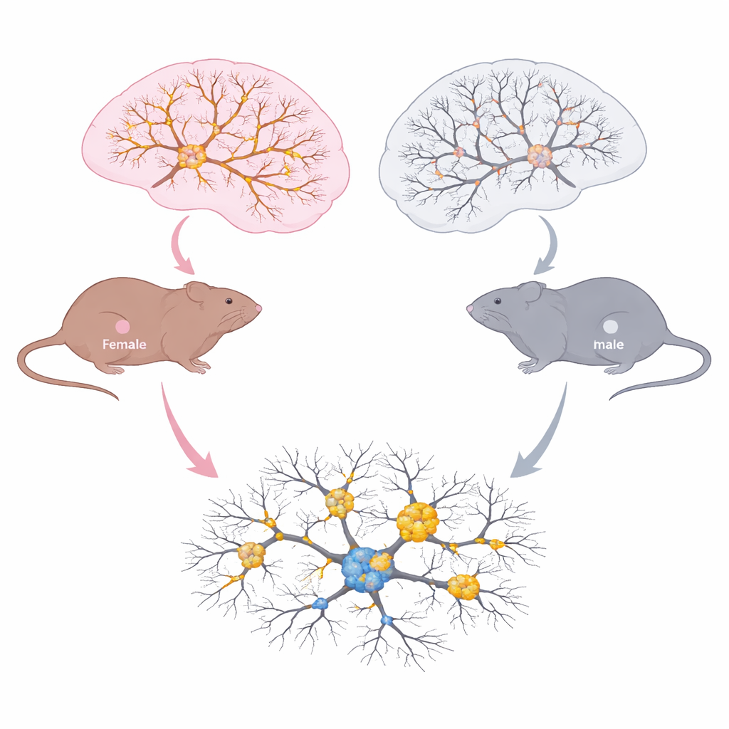
How male and female brains age differently
The team examined brain regions important for memory and decision making at three life stages: young adults, middle age, and older symptomatic animals. In the hippocampus and prefrontal cortex, they measured total tau, phosphorylated tau, acetylated tau at lysine 174, and small tau clusters known as oligomers. Both sexes accumulated more tau oligomers with age, a sign of worsening tauopathy. But the pattern of chemical changes differed sharply. In females, total tau and tau carrying the acetyl mark at lysine 174 rose with age, and this increase tracked with growing tau burden. In males, phosphorylation of tau increased as they aged, but acetylation at lysine 174 did not. Microscopy showed that aged females had more acetylated tau spread through key hippocampal zones and more overlap between acetylated and phosphorylated tau, together with heightened activation of microglia, the brain’s immune cells.
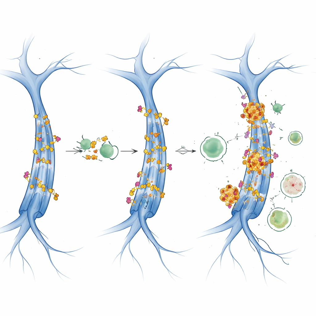
When the brain’s cleaning crews fall behind
To understand why tau built up differently by sex, the researchers probed the brain’s protein maintenance systems. They looked at enzymes that add acetyl marks (notably a pair called CBP/p300), enzymes that remove them (sirtuins), and enzymes that supply the acetyl “fuel” acetyl-CoA. In females, CBP/p300 levels climbed with age, matching the rise in acetylated tau, while acetyl-CoA–producing enzymes and deacetylating sirtuins changed little. This suggests that overactive acetyl-transfer enzymes, rather than fuel supply or removal, drive higher tau acetylation in females. The team also examined autophagy, the cell’s recycling pathway that engulfs and digests unwanted proteins and structures. With aging, several autophagy markers shifted in complex, sex-dependent ways. Females showed signs of reduced formation and flux of the recycling vesicles that help clear tau, while males displayed different patterns, including buildup of a cargo adaptor protein and increased reliance on a related cleanup route.
Signals that slow cellular recycling
The study further explored two master regulators of cell growth and energy use: mTOR and AMPK. When mTOR is highly active, it tends to shut down autophagy, while AMPK promotes it under energy stress. In these tau mice, AMPK activity looked largely stable across age and sex, but mTOR-related signals changed in an age- and sex-specific manner. The data point toward mTOR-driven inhibition of autophagy, intersecting with the acetylation machinery. In females, rising CBP/p300 and acetylated tau coincided with markers of impaired autophagic flux, suggesting a feed-forward loop: more acetylated tau is produced, and at the same time, the system that should dispose of it becomes less efficient, allowing toxic tau clusters to accumulate.
What this means for understanding and treating disease
For non-specialists, the key message is that male and female brains do not handle the same disease protein in the same way as they age. In this humanized tau mouse model, females are especially prone to adding an acetyl tag to tau, driven by higher activity of specific enzymes and accompanied by faltering cellular recycling. Males, in contrast, show more changes in phosphorylation and different alterations in protein cleanup pathways. Although the work was done in mice and with relatively small groups, it supports the idea that acetylation is a critical, sex-sensitive step in tau buildup. That makes acetylation-related enzymes and autophagy regulators attractive targets for treatments tailored to biological sex, with the long-term goal of slowing or preventing tau-driven brain decline.
Citation: Sabir, U., Csubak, B.A., Ilchenko, S. et al. Sex-specific effects of acetylation on tauopathy in aging htau mice. Sci Rep 16, 11862 (2026). https://doi.org/10.1038/s41598-026-41691-0
Keywords: tau acetylation, sex differences, autophagy, Alzheimer’s disease, neurodegeneration