Clear Sky Science · en
Comparing DXA and MRI body composition measurements in cross-sectional and longitudinal cohorts
Why Measuring Fat and Muscle Matters
As we age, our bodies quietly reshuffle how much fat and muscle we carry and where these tissues sit. Those shifts can raise the risk of diabetes, heart disease, and frailty, or signal whether lifestyle changes are really working. Doctors and researchers rely on body-scanning technologies to track these changes, but not all scanners see the body in the same way. This study asks a practical question with big implications: when is a quick, widely used X-ray scan good enough, and when do we need the more detailed, costly MRI scan?

Two Different Ways to Look Inside the Body
The research compares two common whole-body imaging methods. Dual X-ray absorptiometry, often called DXA, uses low-dose X-rays to create a flat, two-dimensional image and then estimates how much of the body is fat and how much is lean tissue. It is fast, relatively inexpensive, and already used in many clinics and large health studies. Magnetic resonance imaging, or MRI, builds a full three-dimensional picture using magnets and radio waves, allowing doctors to distinguish different fat depots and individual muscles in detail. However, MRI scans take longer, cost more, and require specialist analysis, so they are less practical for routine use on very large groups.
A Large-Scale Test in Real People
To find out how closely these methods agree, the authors used data from more than 32,000 adults in the UK Biobank, a long-running health project. Every participant had both DXA and MRI scans on the same day, focused on two key body regions: the "android" area around the abdomen and the "gynoid" area around the hips and thighs. A smaller group of just over 3,000 people returned for another round of scans about two and a half years later, giving the researchers a rare opportunity to compare how each method tracks change over time. The team carefully reproduced the DXA regions on the MRI images so that both tools were measuring the same anatomical areas.
What the Scanners Agree On—and Where They Differ
The good news is that DXA and MRI gave very similar answers for body fat in these regions, including deep belly fat that surrounds internal organs. For overall fat mass and visceral fat, the two techniques lined up closely in both men and women across a wide range of body sizes. The story changed, however, when looking at lean tissue. DXA consistently reported higher lean mass than MRI, particularly in the abdominal region, where DXA estimates were roughly double the MRI values. Part of the reason is that DXA’s "lean" category bundles together muscle with other non-fat tissues such as liver and kidneys, while MRI can separate these structures.
Missed Muscle Loss Over Time
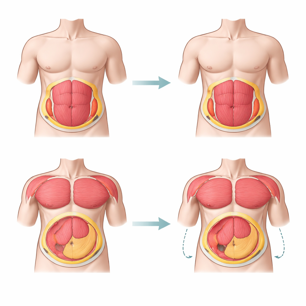
The differences became especially important in the follow-up scans. Over just 2.3 years, MRI detected a 4–5% drop in muscle and lean mass in both abdominal and hip regions for men and women—a pattern consistent with normal age-related muscle loss. DXA, by contrast, showed almost no change in most groups and even suggested a small increase in abdominal lean mass in women. This means that in a relatively short time frame, DXA could fail to spot real declines in muscle, or could misinterpret shifts between muscle and other lean tissues. For studies of ageing, sarcopenia (age-related muscle loss), or subtle effects of diet and exercise, this blind spot could be crucial.
What This Means for Health Studies
For large health surveys and routine checks where the main goal is to estimate body fat and deep belly fat across many people, DXA appears to be a practical and sufficiently accurate choice. However, when the focus is on muscle, or on tracking small changes in body composition over time, this study shows that DXA can give a misleading picture by overestimating lean mass and missing gradual declines. MRI, though more costly and complex, offers a truer, more detailed view of how fat and muscle change with age and lifestyle. In simple terms, DXA is a good wide-angle snapshot for fat, but MRI is the sharper close-up needed when muscle and long-term change really matter.
Citation: Basty, N., Thanaj, M., Whitcher, B. et al. Comparing DXA and MRI body composition measurements in cross-sectional and longitudinal cohorts. Commun Med 6, 227 (2026). https://doi.org/10.1038/s43856-026-01440-w
Keywords: body composition, DXA, MRI, sarcopenia, visceral fat