Clear Sky Science · en
FKBP51 disrupts the insulin signaling pathway and impairs mitochondrial bioenergetics in HepG2 cells
Stress, Sugar, and the Liver
Modern life brings chronic stress, rich diets, and soaring rates of type 2 diabetes. This study looks at a little-known stress‑related protein called FKBP51 and how it changes the way liver cells respond to insulin and use energy. By zooming in on human liver‑derived cells in the lab, the researchers show that FKBP51 can both blunt insulin’s signal and quietly drain energy production inside the cell’s power plants, the mitochondria. Understanding this tug‑of‑war may help explain why long‑term stress and metabolic diseases so often go hand in hand.
A Stress Protein Steps into Metabolism
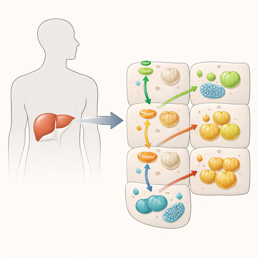
FKBP51 is best known as a helper protein turned on by the stress hormone cortisol. Earlier work linked it to weight gain and insulin resistance in muscle, fat, and brain. However, its role in the liver—an organ central to blood sugar control—was not well understood. In this study, scientists used HepG2 cells, a widely used human liver cell model, and artificially boosted FKBP51 levels to see how this would affect insulin signaling and sugar handling. They focused on key molecules in the insulin pathway and on how mitochondria generate energy, asking whether FKBP51 could be a missing link between stress, liver dysfunction, and high blood sugar.
Insulin Signal Weakened, Yet Sugar Storage Rises
When insulin binds to liver cells, it normally activates a chain of proteins that lower blood sugar by promoting sugar storage as glycogen and reducing new sugar production. The team confirmed that extra FKBP51 weakens this chain: it reduced activation of Akt, a central insulin messenger, and of FOXO1, a regulator of genes that drive the liver to make new glucose. On paper, this should have led to poorer control of sugar. Surprisingly, the opposite happened in some respects. FKBP51‑rich cells made less new glucose even when pushed to do so, and when insulin was present, they actually stored more glycogen than control cells. This suggests that FKBP51 scrambles the usual logic of the insulin pathway, dampening some signals while enhancing others through alternative routes.
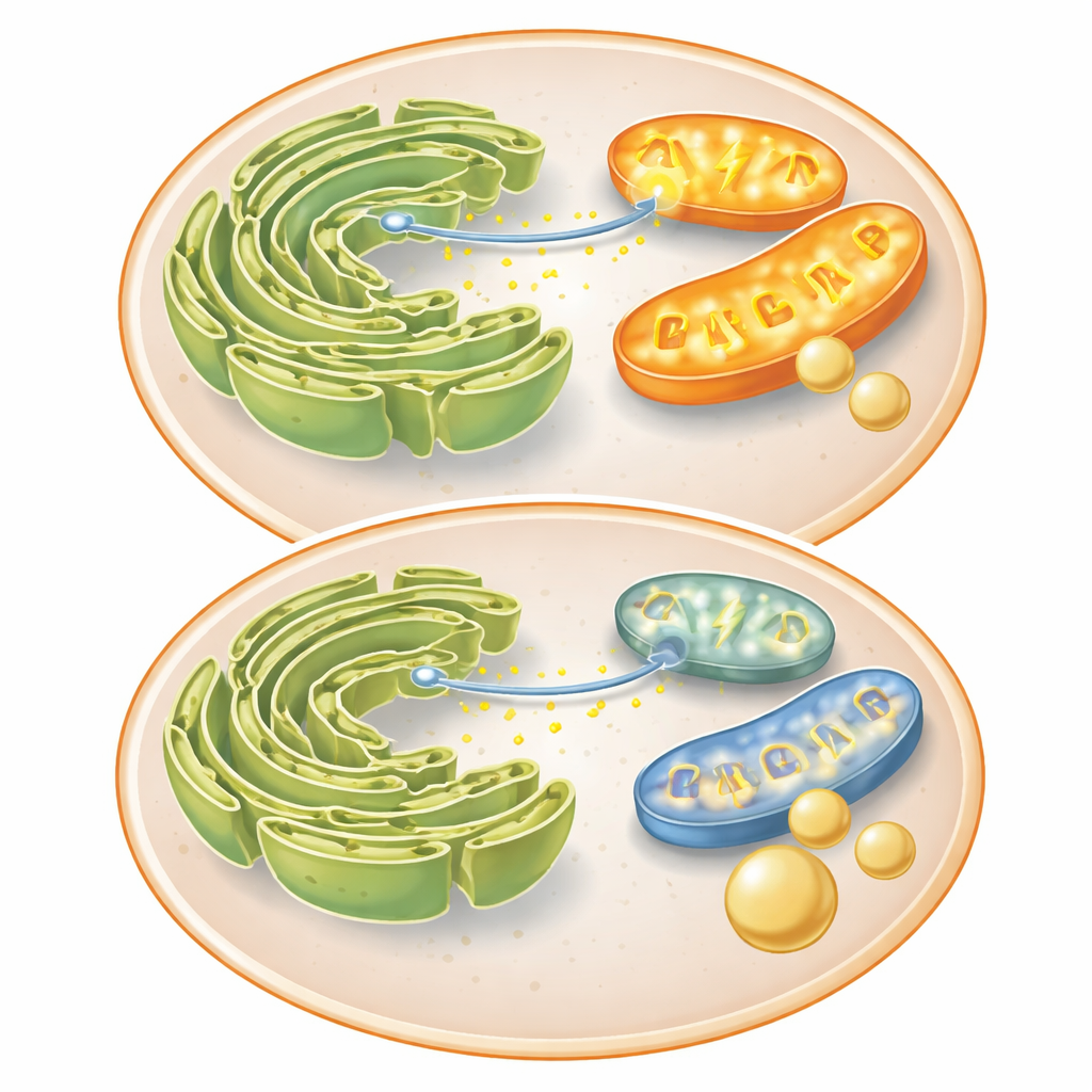
Power Plants Underperform Without Changing Shape
Because the liver’s ability to make or store sugar depends heavily on mitochondria, the researchers next examined these organelles in detail. They found that about half of the FKBP51 signal within cells sits at mitochondria, confirming a physical link. Yet, under the microscope, mitochondrial size and shape looked largely normal, even when FKBP51 was high or insulin was added. Instead, the changes were functional. Both FKBP51 overexpression and insulin treatment reduced how much oxygen mitochondria consumed, lowered their electrical membrane potential, and cut cellular ATP, the chemical currency of energy. Importantly, FKBP51‑rich cells started from an already depressed energetic state, and insulin pushed ATP levels down even further, without a rise in damaging reactive oxygen species.
Broken Conversation Between Two Key Organelles
To understand why mitochondria were underperforming, the team looked at proteins that connect them to another structure, the endoplasmic reticulum (ER), where calcium is stored. The protein Mitofusin 2, important for both mitochondrial fusion and tight contact with the ER, dropped when FKBP51 was high. These contacts normally let calcium move quickly from the ER into mitochondria, boosting energy‑producing enzymes. In control cells, a chemical signal caused a strong burst of calcium into mitochondria; insulin and FKBP51 both slowed this transfer. FKBP51 also reduced calcium spikes in the cell fluid itself, implying that it hampers calcium release from the ER. With less calcium reaching mitochondria, their energy output falls, nudging the cell away from costly processes like glucose production and toward storing incoming sugar instead.
What This Means for Health
Put simply, the study suggests that FKBP51 acts as a double‑edged stress responder in liver cells. On one side, it interferes with insulin’s early signaling steps, a feature associated with insulin resistance. On the other, it weakens the energy‑making machinery by disturbing calcium exchange between the ER and mitochondria. The combined effect is a liver cell that makes less new glucose but is more inclined to stash available sugar as glycogen, even when classic insulin signals are partially muted. For people, this work hints that chronic stress—through proteins like FKBP51—may reshape liver energy use in subtle ways that contribute to metabolic disease. Targeting FKBP51 or the ER‑mitochondria calcium connection could one day offer new strategies to fine‑tune blood sugar control without overtaxing the cell’s power plants.
Citation: Donoso-Barraza, C., Díaz-Roblero, M., Sepúlveda, C. et al. FKBP51 disrupts the insulin signaling pathway and impairs mitochondrial bioenergetics in HepG2 cells. Sci Rep 16, 9896 (2026). https://doi.org/10.1038/s41598-026-40414-9
Keywords: insulin resistance, liver metabolism, mitochondrial function, cellular stress, calcium signaling