Clear Sky Science · en
A geometric morphometrics approach to sex estimation of infants from 0 to 6 years using the auricular surface
Why Tiny Bones Matter for Big Human Stories
Archaeologists and forensic scientists often work with bones to reconstruct how people lived, died, and were treated by their communities. Yet when it comes to babies and very young children, one crucial piece of information is frequently missing: whether the child was biologically male or female. This study explores whether subtle shape differences in a small joint surface of the hip bone in infants—from birth to six years old—can help estimate sex, potentially opening a new window onto childhood in past populations.
Looking for Clues in the Infant Hip
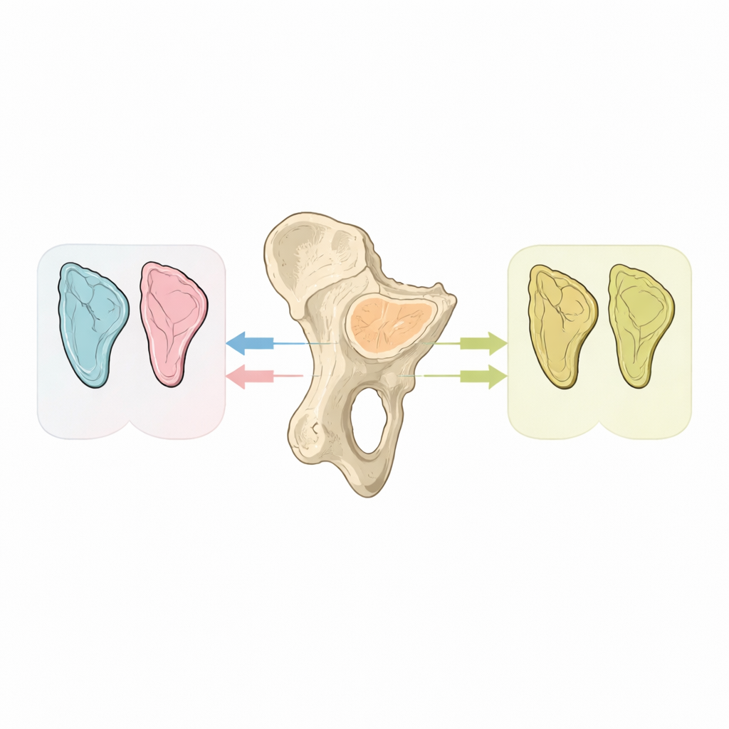
The researchers focused on a specific part of the hip bone called the auricular surface, where the spine connects to the pelvis. Earlier work suggested this area might differ between males and females even before puberty, but results were mixed. To test this more rigorously, the team used a modern skeletal collection from Lisbon, Portugal, where the age and sex of each individual were already known. They selected 46 infants and young children, aged from birth to just under seven years, and concentrated on one side of the pelvis for consistency.

Turning Bones into 3D Data
Instead of taking simple measurements with calipers, the team created detailed three-dimensional digital models of each hip bone using a portable surface scanner. On these virtual bones, they mapped a series of carefully chosen points around the outline of the auricular surface. Some points marked clear anatomical corners, while others were automatically spread along curves to capture smooth changes in shape. Using a set of mathematical tools known as geometric morphometrics, they could then compare the size and shape of this joint surface across individuals in a precise, standardized way.
What Changes with Age and What Does Not
When the researchers looked at size, they found that the auricular surface generally grew larger with age, but the growth patterns for boys and girls shifted over time. Under one year old, the joint surface was similar in size for both sexes, with a slightly higher median for girls. Between one and almost four years, girls tended to have larger joint surfaces, while boys pulled ahead in size between four and almost seven years. However, these differences were modest and did not reach the level scientists usually require to call them statistically significant, meaning they cannot yet be relied on for sex estimation.
Subtle Shape Signals in the Youngest Infants
The team then turned to shape, asking whether the outline of the auricular surface differed between males and females in distinct age groups. For the overall sample, there was no clear separation between the sexes. But when they looked only at infants under one year, the picture became more intriguing. Statistical tests did not quite meet the standard threshold for significance, largely because there were only seven infants in this age band. Nonetheless, visual inspection of the results showed that male and female infants tended to cluster in different regions of shape space, hinting at a real underlying difference in how this joint surface is formed in the first months of life.
Hormones, First Steps, and Vanishing Differences
The authors suggest that these early shape differences may reflect a brief surge of sex hormones that occurs in infancy, sometimes called “minipuberty.” In boys, testosterone levels rise soon after birth and fall back within six to nine months; in girls, estrogen levels are elevated and then decline by about two years of age. This hormonal window may temporarily push male and female bones along slightly different developmental paths. As children begin to stand and walk—usually around their first birthday—the mechanical forces of bipedal movement become more similar between the sexes, possibly smoothing out these early differences so that the auricular surface looks alike in older boys and girls.
What This Means for Studying the Past
Overall, the study shows that high-resolution 3D analysis of the auricular surface is promising but not yet reliable as a stand-alone method for determining the sex of infants. There are hints of meaningful differences in babies under one year, but the sample is too small and the patterns too subtle to use confidently in real-world forensic or archaeological cases. The authors recommend expanding the number and diversity of skeletons studied, examining other features of the hip bone alongside the auricular surface, and exploring artificial intelligence approaches that might detect complex combinations of traits invisible to the naked eye. If refined, such methods could eventually help researchers reveal the often-hidden lives of boys and girls in past societies, from nutrition and disease to burial customs and social care.
Citation: Simão, P., Garcia, S.J. & Godinho, R.M. A geometric morphometrics approach to sex estimation of infants from 0 to 6 years using the auricular surface. Sci Rep 16, 11422 (2026). https://doi.org/10.1038/s41598-026-35321-y
Keywords: infant skeletal sex estimation, auricular surface, geometric morphometrics, bioarchaeology, pelvis development