Clear Sky Science · en
“Micro-managing” immune activation and protein turnover: microglial lysosomes in the context of health and disease
Watching the Brain’s Cleanup Crew
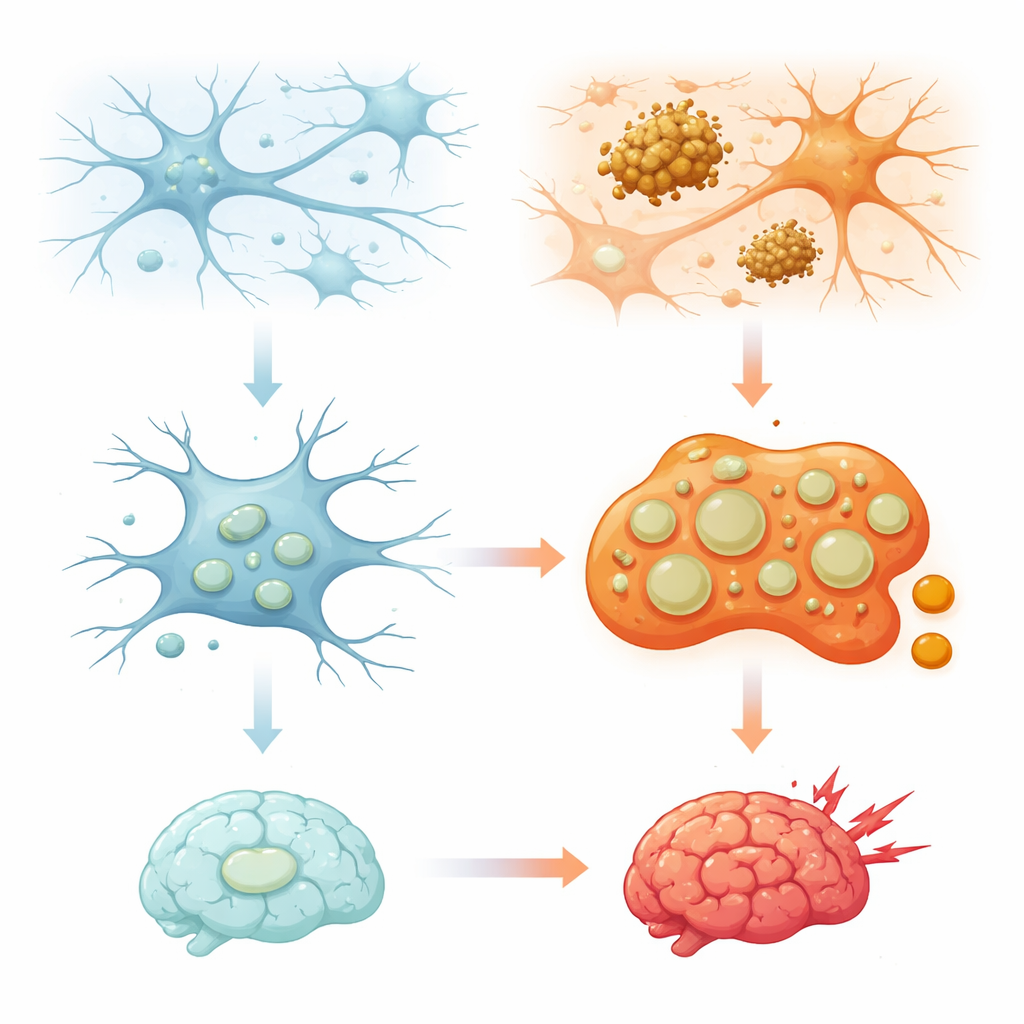
Deep inside the brain, tiny immune cells called microglia act as janitors and first responders. They swallow germs, dead cells, and clumps of misfolded proteins that can build up with age or in diseases like Alzheimer’s. This review article explores a less familiar part of that story: the role of microglial lysosomes, the cell’s internal “recycling centers.” By tracing how these compartments are controlled, how they change with stress and aging, and how they differ across cell types, the authors show why fine‑tuning microglial lysosomes could be key to preventing or treating neurodegenerative diseases.
How Brain Janitors Use Their Inner Recycling Bins
Lysosomes have long been seen as simple trash cans that break down worn‑out proteins and organelles. The past decade has revealed a more complex picture: lysosomes also sense nutrients, help control energy use, shape immune responses, and even take part in cell death decisions. In microglia, which constantly patrol the brain, these compartments are especially important because they handle large amounts of swallowed material, from microbes to protein aggregates and synapses that need pruning. The review explains that microglial lysosomes are not all alike; they form different subpopulations whose composition, position inside the cell, and behavior shift with stress, aging, and disease. Understanding how this diversity is built and maintained is essential to grasping why some brain regions or cell types are more vulnerable than others.
Switches That Turn Lysosomes Up or Down
At the genetic level, microglial lysosomes are controlled by a network of master switches, particularly a family of transcription factors that includes TFEB. When cells are stressed or overloaded, TFEB moves into the nucleus and switches on a set of genes that build more lysosomes and boost their activity. Studies in zebrafish and mouse models show that this TFEB‑driven program is crucial for microglia to migrate properly, acidify their lysosomes, and mount a strong immune response against protein aggregates such as toxic forms of Tau. Other regulators push in the opposite direction. For example, an overactive Parkinson’s‑linked enzyme, LRRK2, keeps these transcription factors out of the nucleus in microglia and macrophages, weakening lysosomal breakdown and potentially raising disease risk. Together, these findings highlight a tug‑of‑war between pathways that enhance lysosomal function under stress and those that restrain it to maintain everyday balance.
From Protective Cleanup to Harmful Inflammation
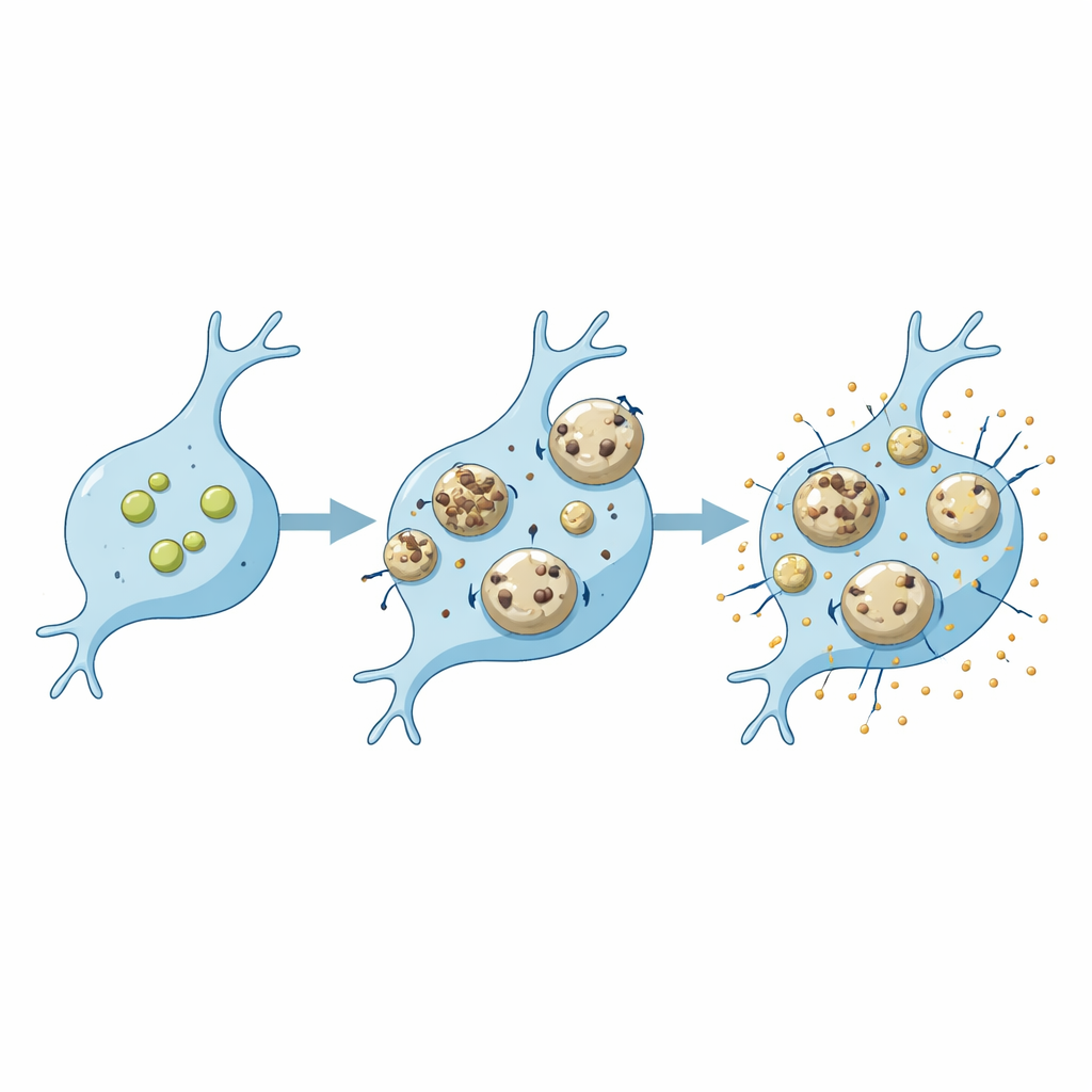
Microglial lysosomes sit at the crossroads of waste removal and inflammation. When microglia engulf immune‑stimulating substances—such as bacterial components, amyloid‑beta, or other protein aggregates—their lysosomes not only degrade this material but also help activate and release inflammatory signals. Proteases called cathepsins, normally confined within lysosomes, play central roles: they help convert inactive immune messengers into active ones, promote the release of inflammatory molecules, and, when leaked or secreted, can even damage nearby neurons. Another lysosomal protein, CD68, is widely used as a marker for active, phagocytic microglia and rises with aging, oxidative stress, and Alzheimer’s pathology. Yet its exact function in clearance and inflammation remains unclear. Importantly, drugs that restore lysosomal acidity or antibodies that engage microglial clearance pathways can calm inflammation and improve protein removal in animal models, hinting at new therapeutic routes.
Different Cells, Different Vulnerabilities
The review emphasizes that the same genetic defect can affect microglial and neuronal lysosomes in distinct ways. Loss of proteins such as progranulin, SORLA, or C9ORF72, all linked to dementia or motor neuron disease, leads to one pattern of lysosomal changes in microglia and another in neurons. In microglia, these changes often alter how well they degrade cargo like amyloid‑beta or synaptic debris and how strongly they respond with inflammatory signals. Lysosomes are also prone to physical damage when they are packed with sticky aggregates like amyloid‑beta or alpha‑synuclein. In microglia, such damage can trigger protective repair pathways and a form of targeted self‑cleaning called lysophagy; if these safeguards fail, leaked enzymes can drive cell death and further inflammation. Evidence suggests that microglia may have more robust repair options than neurons, but how this changes with age or disease is still being investigated.
Hidden Diversity Inside Microglial Cells
Beyond differences between cell types, microglia show striking variation among themselves and even within a single cell. Work in aging brains of mice and monkeys has uncovered two broad classes of microglia distinguished by the amount of autofluorescent, lysosome‑rich storage material they accumulate. One subset builds up complex storage bodies and high levels of lysosomal proteins and enzymes over time, then declines in number at very old ages. Within individual microglia, lysosomes in the long branching processes may differ from those in the cell body, reflecting local demands for pruning synapses or handling myelin debris. The authors outline open questions: how resting and activated lysosomes differ, whether activation sparks the birth of new lysosomes, how CD68‑positive lysosomes are specialized, and whether some lysosomal pools are dedicated to secretion while others focus on digestion. Answering these questions will require advanced imaging and molecular tools but promises to deepen our understanding of brain health.
Why These Tiny Compartments Matter for Brain Health
Altogether, the review portrays microglial lysosomes as finely tuned hubs where cleanup, metabolism, and immunity intersect. When these compartments work well, they help microglia quietly maintain brain circuits by clearing debris and curbing threats. When they are overtaxed, genetically impaired, or physically damaged by toxic aggregates, they can flip from protective recycling centers into sources of chronic inflammation and neuronal injury. By mapping the regulatory networks, damage responses, and hidden diversity of microglial lysosomes, the authors argue that future therapies might selectively boost or rebalance these systems—enhancing beneficial clearance while limiting harmful inflammation—to slow or prevent neurodegenerative diseases.
Citation: Somodji, E.A., Gowrishankar, S. “Micro-managing” immune activation and protein turnover: microglial lysosomes in the context of health and disease. npj Dement. 2, 35 (2026). https://doi.org/10.1038/s44400-026-00086-8
Keywords: microglia, lysosomes, neuroinflammation, Alzheimer’s disease, protein aggregation