Clear Sky Science · en
Clinical and histopathological characterization of metastatic lobular breast cancer: lessons learned from post-mortem tissue donation programs
Why this matters for people living with breast cancer
When breast cancer spreads through the body, doctors typically rely on a single metastatic biopsy and routine scans to choose treatment. This study shows that, for a common subtype called lobular breast cancer, that approach may miss important differences between tumors and even miss entire hidden deposits of cancer. By studying tissue generously donated after death, researchers were able to map this disease in unprecedented detail and reveal why some patients may not be getting the most tailored care possible.
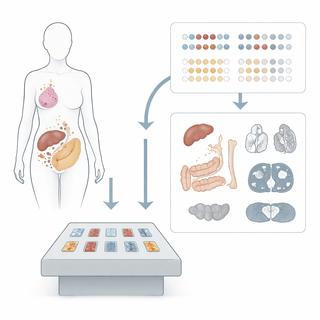
Looking closely at a quiet but widespread cancer
Invasive lobular carcinoma is the second most common form of breast cancer. Unlike more familiar breast cancers that form solid lumps, lobular tumors spread as single, scattered cells that can be hard to see on scans. They also have a habit of turning up in unusual places, such as the stomach, intestines, and female reproductive organs, in addition to more typical sites like liver and bone. Because biopsies during life are limited by what is safe and practical to sample, much about the true extent and biology of metastatic lobular cancer has remained unknown.
A unique view made possible by tissue donation
To overcome these limits, two rapid post-mortem donation programs in Belgium and the United States collected tissue from 12 people who died with metastatic lobular breast cancer. Within a few hours after death, doctors systematically sampled visible and randomly chosen organs, ultimately examining 306 metastatic deposits—about 27 per patient. They compared these spots with each person’s original breast tumor, measuring standard markers used worldwide to guide therapy: hormone receptors (estrogen and progesterone), the HER2 protein targeted by several drugs, a growth marker called KI67, and the presence of immune cells in and around the tumors.
Many metastases, many different tumor profiles
The results revealed striking differences between metastases, even within the same person. Although most original tumors were strongly hormone-receptor positive, over half of the patients had some metastases that had partly or completely lost estrogen or progesterone receptors. Overall, hormone levels in metastases were significantly lower than in the primary tumor, while the growth marker KI67 tended to be higher, suggesting more aggressive behavior at distant sites. HER2 remained formally “negative” in most primaries, but nearly every patient had at least some metastases with low or ultra-low HER2 protein levels—enough to potentially qualify for newer HER2-targeted drugs. At the same time, immune cells were generally scarce in both primary tumors and metastases, reinforcing the idea that many lobular cancers are poorly inflamed and may respond less well to immunotherapy.
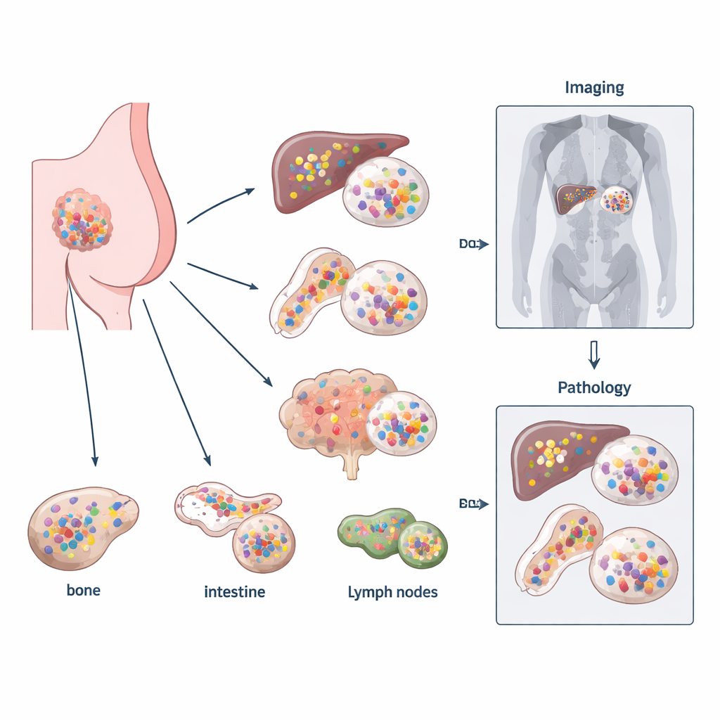
Scans versus the microscope: what gets missed
The team also compared the last CT or whole-body MRI scans obtained before death with what was actually found under the microscope at autopsy in nine patients. Overall, imaging and pathology agreed on organ involvement about three-quarters of the time. But important mismatches emerged. For instance, the liver sometimes looked normal on scans yet harbored microscopic lobular metastases. In other cases, imaging suggested spread to organs like the uterus or intestines that could not be confirmed in sampled tissue. These discrepancies highlight both the difficulty of detecting the subtle growth pattern of lobular cancer on standard imaging and the risk of missing scattered tumor cells if tissue sampling during life is limited.
What this means for patients and care teams
Together, these findings show that metastatic lobular breast cancer is more varied and widespread than a single biopsy or routine scan can capture. A metastasis sampled from one site may not reflect what is happening elsewhere in the body, particularly with respect to hormone and HER2 status that directly determine treatment options. The study suggests that repeating biomarker testing whenever a new metastatic site is sampled, and developing better whole-body tools such as advanced imaging or blood-based tests, will be crucial to match patients with the best therapies—including newer drugs for HER2-low disease. Thanks to the patients who chose tissue donation, clinicians now have a clearer picture of this elusive cancer and a roadmap for improving how it is detected and treated.
Citation: Zels, G., Van Baelen, K., Chang, A.C. et al. Clinical and histopathological characterization of metastatic lobular breast cancer: lessons learned from post-mortem tissue donation programs. npj Breast Cancer 12, 48 (2026). https://doi.org/10.1038/s41523-026-00912-5
Keywords: metastatic lobular breast cancer, tumor heterogeneity, post-mortem tissue donation, HER2-low disease, cancer imaging