Clear Sky Science · en
Identifying GFAP-expressing cell susceptibility to SARS-CoV-2 infection using human iPSC-derived neural cells
How a COVID-19 Virus Can Reach Brain Cells
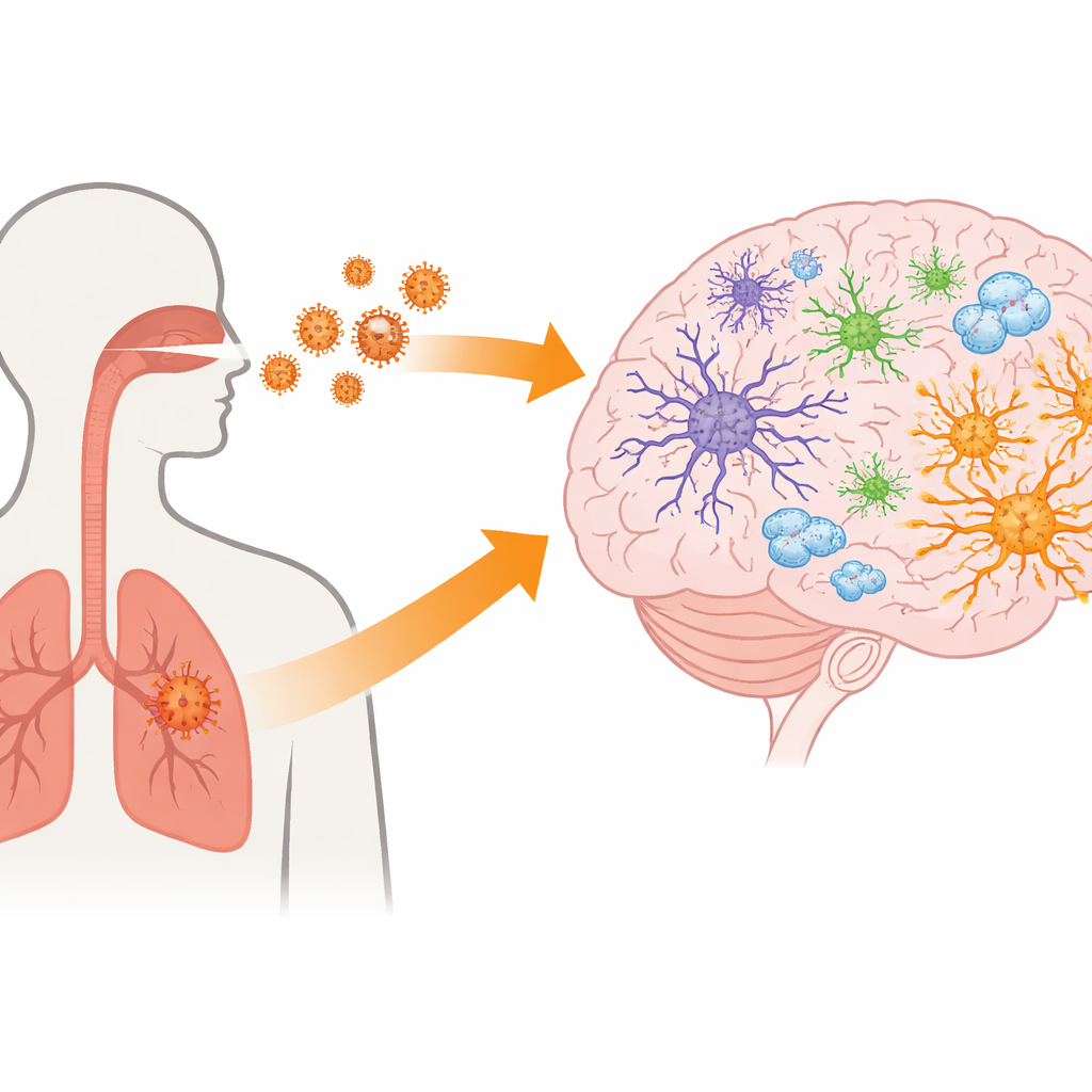
Many people with COVID-19 report problems such as headaches, loss of smell, confusion, or lingering “brain fog.” These symptoms suggest that the virus that causes COVID-19, SARS-CoV-2, may in some cases affect the brain. Yet it has been difficult to study exactly which brain cells are at risk and by what route the virus gains a foothold. This study builds a fast, human-based laboratory model of brain tissue to ask a simple but important question: which brain cells are most vulnerable to SARS-CoV-2, and what might explain that vulnerability?
Building a Mini Brain Cell Sheet in a Dish
The researchers started with human induced pluripotent stem cells, which are adult cells reprogrammed back into a flexible, embryo-like state. Using a stepwise recipe of growth factors and nutrients, they encouraged these cells to mature over just two weeks into a flat, two-dimensional layer of brainlike cells. Careful gene “fingerprinting” at the single-cell level showed that this sheet contained a mix of cell types typically found in the outer layer of the brain, including several kinds of nerve cells and support cells. Remarkably, the variety of cell types after only 14 days resembled that seen in much older, three-dimensional brain organoids that usually take many months to grow.
Matching a Faster Model to Complex Brain Organoids
To judge how realistic their new system was, the team compared its gene activity patterns to those from previously published brain organoids grown for six and ten months. Both systems shared many of the same major cell groups, such as radial glia (immature support cells), excitatory and inhibitory neurons, and early-stage neurons. One key difference was that the quick 2D model contained more interneurons and astrocytes, a type of star-shaped helper cell, than the older organoids. This trade-off suggests that while the 2D cultures lack the full 3D architecture of brain tissue, they capture a rich mix of cell identities in a fraction of the time, making them especially useful for rapid studies during emerging outbreaks.
Tracking Which Brain Cells the Virus Prefers
The team then exposed these mixed brain cell cultures to different strains of SARS-CoV-2, including the original Wuhan virus and an Alpha variant, and watched what happened over the next three days. The virus could infect the 2D neural cultures and produce new viral particles, whereas a commonly used nerve cell line (SH-SY5Y) did not support infection. Even though fewer than 2 percent of the cells showed signs of infection, the amount of virus released into the culture fluid was substantial. When the scientists stained for viral proteins along with markers for different brain cell types, they found that infection landed mainly in cells that made a protein called GFAP, characteristic of astrocyte-like cells, rather than in neurons.
Looking for the Viral Doorway on Astrocytes
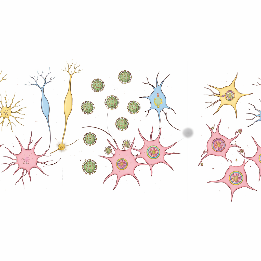
SARS-CoV-2 usually enters cells by binding to a protein called ACE2, often helped by other factors on the cell surface. Surprisingly, when the researchers measured ACE2 and related entry factors across the whole culture, levels were very low, and there was little difference between the susceptible 2D brain cells and the resistant SH-SY5Y cells. Only about 2 percent of cells showed detectable ACE2 protein, and these did not overlap with GFAP-positive astrocyte-like cells. To get a clearer view, the team returned to single-cell gene data and examined a broader panel of candidate entry genes. Here, astrocyte clusters stood out for much higher levels of a gene called BSG, which encodes the surface protein CD147, as well as HSPA5, another proposed helper for viral entry. This pattern suggests that, at least in this model, SARS-CoV-2 may exploit alternative doorways on astrocyte-like cells rather than relying on ACE2.
What This Means for the Brain and COVID-19
In this streamlined human brain cell model, SARS-CoV-2 can infect cells but focuses mainly on astrocyte-like cells that carry high levels of BSG, while leaving most neurons untouched and without triggering a strong, widespread inflammatory reaction. For non-specialists, the takeaway is that the virus appears able to reach and infect certain support cells in the brain, and that an alternate surface protein, CD147 (from the BSG gene), may help open the door. The model does not yet capture all features of the living brain, such as blood vessels or immune cells, but it offers a rapid and realistic way to probe how COVID-19 might disturb brain function and to test ideas about long COVID and other lingering neurological effects.
Citation: Asavapanumas, N., Chaiwijit, P., Suksatu, A. et al. Identifying GFAP-expressing cell susceptibility to SARS-CoV-2 infection using human iPSC-derived neural cells. Sci Rep 16, 10433 (2026). https://doi.org/10.1038/s41598-026-41177-z
Keywords: COVID-19 and brain, SARS-CoV-2 neurotropism, astrocytes, brain organoids and iPSCs, viral entry receptors