Clear Sky Science · en
Intravitreal photoswitch therapy in advanced retinitis pigmentosa: a phase 1 open-label trial
A new ray of hope for fading sight
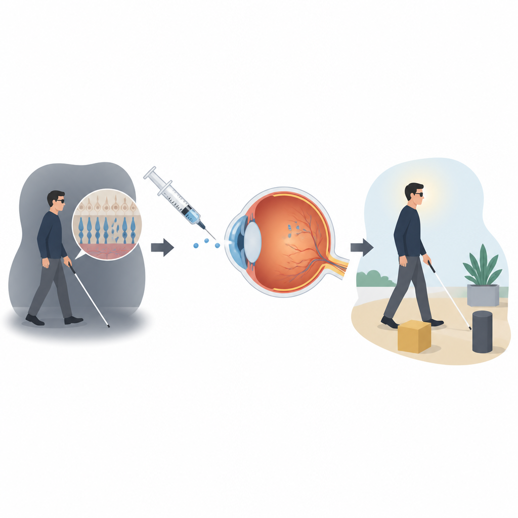
Retinitis pigmentosa is a group of inherited eye diseases that slowly narrow and dim a person’s vision, often leading to near or complete blindness by midlife. For the many people affected, current treatments can only slow damage at best and usually cannot bring lost sight back. This study reports an early clinical test of a different idea: using a light sensitive drug, delivered into the eye, to make surviving nerve cells respond to light again and possibly restore simple visual signals.
Turning eye nerve cells into light sensors
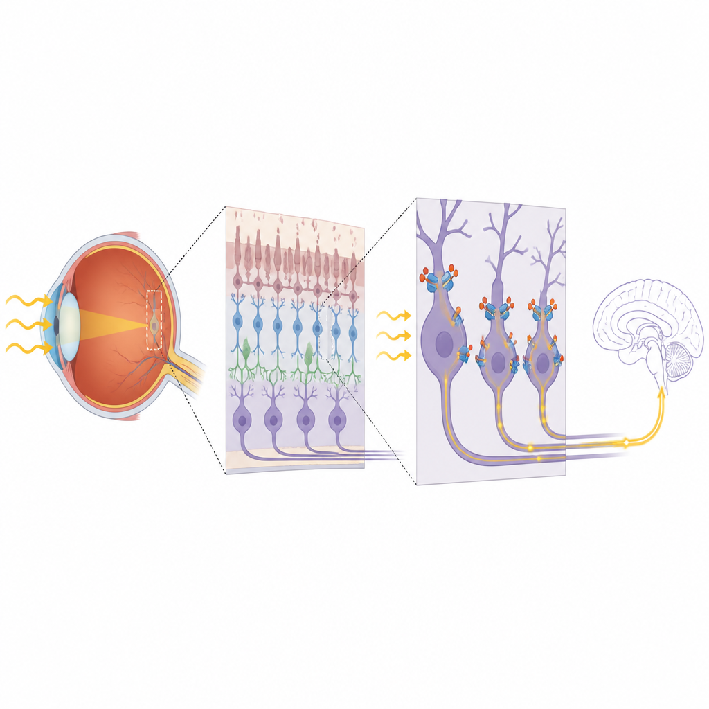
In healthy eyes, light is first caught by rods and cones, the classic “photo cells” of the retina. In advanced retinitis pigmentosa, most rods and cones have died, but deeper layers of nerve cells, including retinal ganglion cells that send signals to the brain, can remain. The researchers tested a small molecule called KIO-301, built to slip into these surviving nerve cells and change shape when exposed to light. In animal studies, this light driven shape shift allowed the drug to briefly block and unblock tiny pores in the cell membrane, turning incoming light into electrical activity that could travel along the optic nerve, bypassing the missing rods and cones.
First steps in people with very low vision
The ABACUS-1 trial was a first in human, phase 1 safety study carried out in Australia. Six adults with severe vision loss from retinitis pigmentosa took part; some could barely sense light, while others could only detect hand motion or count fingers. Each participant received an injection of KIO-301 into the gel filled space inside one eye, followed later by a higher dose in the other eye if no safety problems appeared. Doctors then followed the participants closely for a month after each injection, checking eye health, general health and how well they could perform a range of simple vision and navigation tasks.
Safety findings and early signs of activity
The main goal was to see whether the drug and the injection procedure were safe in this fragile group. Across all 12 treated eyes and three dose levels, no serious side effects or dose limiting toxicities occurred. A few mild issues, such as brief eye discomfort, eyelid swelling and a small rise in eye pressure in one person, were consistent with known effects of eye injections and resolved with standard care. Importantly, doctors saw no signs of drug related inflammation, scarring or swelling inside the eye on detailed imaging tests. Blood tests showed almost no detectable drug in the circulation, suggesting that the treatment stayed local to the eye.
What patients could see and what the brain showed
Although the study was not designed to prove that vision improved, the team explored several measures that might hint at drug activity. Some participants who had long been unable to see any light reported new, faint perceptions of brightness within a few days of treatment. In controlled tests of walking direction, finding a window or locating a room exit, several treated eyes performed better at early visits than at baseline, with performance tending to peak in the first two weeks and then drift back toward starting levels by day 30. Brain scans using functional MRI added another piece: soon after dosing, flashing visual patterns presented to the treated eye triggered blood flow changes in visual areas at the back of the brain, again strongest in the first two to three days and fading later.
Limits of the trial and what comes next
The authors stress that this was a very small, open label safety trial without a comparison group, and it lasted only a short time. Because of this, they cannot say whether the drug truly improved vision, how strong any benefit might be, or how long it could last. Light intensity during testing was chosen cautiously rather than to maximize effect, and the exact way KIO-301 enters human retinal cells and alters their behavior is still being worked out. Even so, the aligned reports of light sensations, better performance on simple tasks and brain responses to visual stimulation suggest that the drug did affect visual pathways, at least briefly.
A cautious but encouraging beginning
For people living with advanced retinitis pigmentosa, these findings do not yet offer a treatment they can rely on, but they do mark an important milestone. KIO-301, a light sensitive small molecule injected into the eye, appeared safe over the first month in this group and showed early signs that it might help remaining retinal nerve cells pass light driven signals to the brain. This work lays the groundwork for larger, longer studies to test repeated dosing, fine tune light stimulation and determine whether photoswitch therapy can meaningfully improve daily vision in those who have lost so much of it.
Citation: Casson, R.J., Daniels, E., Barras, C.D. et al. Intravitreal photoswitch therapy in advanced retinitis pigmentosa: a phase 1 open-label trial. Nat Med 32, 1865–1870 (2026). https://doi.org/10.1038/s41591-026-04317-6
Keywords: retinitis pigmentosa, vision restoration, photoswitch therapy, retinal ganglion cells, intravitreal injection