Clear Sky Science · en
EpCAM supports exit from pluripotency of embryonic stem cells via Eomes
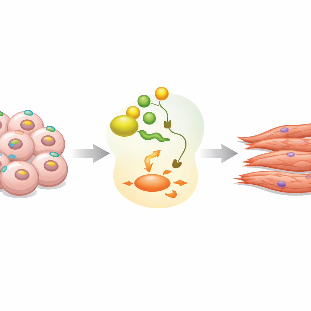
From Blank-Slate Cells to Beating Heart Tissue
Early in development, a tiny clump of embryonic cells has the remarkable ability to turn into any tissue in the body. This study explores how such "blank-slate" mouse stem cells decide to leave that flexible state and commit to becoming heart muscle. The researchers focus on a surface molecule called EpCAM and uncover how it helps flip a key internal genetic switch, Eomes, guiding cells out of pluripotency and toward beating cardiomyocytes. Understanding this control system may improve stem-cell-based strategies for repairing the heart and clarifies how early embryos organize their first tissues.
A Surface Marker with a Hidden Agenda
EpCAM is best known as a marker found on many cancers and on immature stem cells, but its exact role during early development has been unclear. In mouse embryonic stem cells grown as three-dimensional clusters called embryoid bodies, the team observed that EpCAM appears on almost all cells in the initial pluripotent state. As differentiation begins, EpCAM levels briefly rise and then sharply fall in many cells, mirroring patterns seen in real mouse and human embryos. Earlier work had shown that too much or too little EpCAM disrupts normal development, hinting that its timing and location must be precisely controlled.
What Happens When EpCAM Is Missing?
To probe EpCAM’s function, the researchers used CRISPR to create mouse stem cell lines lacking the Epcam gene. When these modified cells were allowed to differentiate without added growth factors, they formed embryoid bodies that rarely produced contracting heart muscle. Markers of cardiomyocytes, such as alpha-cardiac actin, were strongly reduced. Re-introducing EpCAM in a controlled, time-limited way largely restored the appearance of beating areas and heart-specific genes, but only when the pattern of expression mimicked the normal early pulse followed by loss. This showed that EpCAM is not just a passive label: it actively supports the cells’ exit from pluripotency and their ability to become heart tissue.
Linking EpCAM to Internal Gene Switches
Using bulk and single-cell RNA sequencing across several days of differentiation, the team compared normal and EpCAM-deficient cells. They found that, shortly after differentiation starts, cells without EpCAM fail to properly activate a suite of lineage-defining transcription factors, including Eomes, Foxa2, and Gata6. Among these, Eomes emerged as a central player: it is known to drive formation of early mesoderm and definitive endoderm, the tissues that give rise to structures such as the heart and gut. In normal cells, Epcam and Eomes are co-expressed in epiblast and primitive streak–like cells and in early mesoderm, both in the embryoid bodies and in public datasets from real mouse and human embryos. In EpCAM knockouts, this coordinated rise of Eomes is blunted, and pluripotency factors like Nanog linger longer than they should.
A Signaling Relay: EpCAM, Wnt, and Eomes
To uncover how a surface protein like EpCAM can influence an internal regulator such as Eomes, the researchers examined major signaling pathways. Gene-set analyses pointed strongly to Wnt signaling, a pathway well known for shaping early body axes and heart formation. In normal cells, Wnt activity increases as cells traverse the primitive streak toward cardiac fates, in step with EpCAM and Eomes. In EpCAM-deficient cells, this surge is muted. Pharmacologically boosting Wnt-related activity during the earliest days of differentiation partially rescued the formation of beating cardiomyocytes and restored heart-gene expression. In reporter experiments, re-expressing EpCAM directly enhanced Eomes promoter activity, and blocking Wnt reduced this effect, supporting a model in which EpCAM helps drive Eomes through Wnt-dependent routes.
Rescuing the Heart Pathway and What It Means
Finally, the authors asked whether turning Eomes back on is sufficient to bypass the loss of EpCAM. When they transiently re-expressed Eomes in EpCAM-null stem cells, cardiomyocyte formation and contraction rates dramatically improved, closely approaching those of normal cells. In contrast, restoring Foxa2 or Gata6 alone did not reliably fix the defect. Together, these results position EpCAM as an upstream regulator that, at the right moment, boosts Wnt signaling and Eomes, allowing stem cells to shut down their flexible, pluripotent program and commit to heart muscle. For a general reader, the takeaway is that a molecule long viewed as a simple tag on the cell surface actually behaves like a timing device: it helps tell early embryonic cells when to stop being "anything" and start becoming "something"—in this case, the beating cells of the heart.
Citation: Gong, N., Gouda, M., Balaz, A.M. et al. EpCAM supports exit from pluripotency of embryonic stem cells via Eomes. Cell Death Dis 17, 389 (2026). https://doi.org/10.1038/s41419-026-08734-w
Keywords: embryonic stem cells, EpCAM, Wnt signaling, Eomes, cardiomyocyte differentiation