Clear Sky Science · en
The protein kinase DYRK1B is a p53 target gene and functions as a negative feedback regulator of the transcription factor RFX7
Why this matters for cancer treatment
Most cancers carry damage to the famous “guardian of the genome,” a protein called p53. This guardian normally helps cells pause, repair their DNA, or self-destruct when things go wrong. The study summarized here uncovers a new way in which cancer cells can blunt this protective system through another protein, a kinase called DYRK1B, and shows that blocking DYRK1B could make tumor cells more vulnerable to chemotherapy.
A built-in alarm system in our cells
When cells experience stress, such as DNA damage or problems making new cellular components, p53 switches on and launches an emergency program. It does this largely by turning other genes on or off. Some of those genes directly halt the cell cycle or trigger cell death, but p53 also works through additional control proteins that themselves regulate many downstream genes. One of these is RFX7, a transcription factor that has recently emerged as an important tumor suppressor. RFX7 helps activate a network of genes that restrain tumor growth and is often disrupted or muted in human cancers.
A survival-promoting kinase in the spotlight
DYRK1B is an enzyme that adds phosphate groups to other proteins, thereby changing their behavior. Earlier work showed that DYRK1B helps cancer cells endure tough conditions, keep a low-activity state, and repair DNA damage. It is frequently found at abnormally high levels in several solid tumors, and blocking it can make cancer cells more sensitive to chemotherapy or radiation in experimental models. Yet, compared with many other cancer-related enzymes, DYRK1B has remained poorly understood, earning the label of a “dark kinase.” The new study set out to clarify how DYRK1B is controlled and how it fits into the broader stress-response circuitry governed by p53.
From p53 to RFX7 to DYRK1B
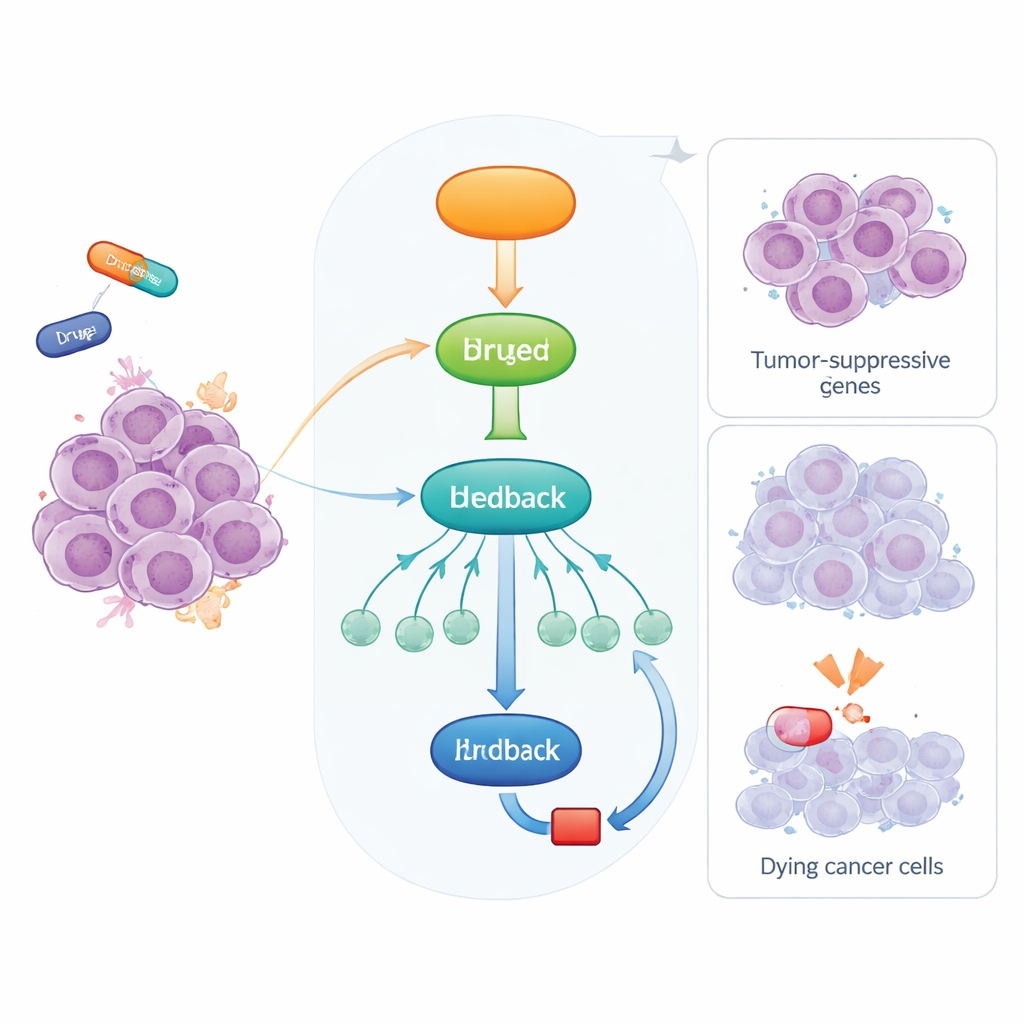
The researchers treated various cancer cell lines with two chemotherapy drugs, doxorubicin and actinomycin D, both of which activate p53. They observed that DYRK1B levels rose strongly after treatment, whereas its close relative DYRK1A did not. Using a drug called Nutlin-3a, which turns on p53 without causing DNA damage, they confirmed that DYRK1B is induced whenever p53 is activated. When p53 was genetically removed, this rise in DYRK1B disappeared, and analysis of tumor data from patients showed that DYRK1B expression tends to track with p53 levels across many cancer types. The team then showed that this induction is indirect: p53 first stimulates RFX7, and RFX7 in turn boosts DYRK1B. Knocking out RFX7 or disabling its ability to enter the nucleus sharply reduced DYRK1B induction, and DYRK1B levels rose at the RNA as well as protein level, confirming genuine gene activation.
A molecular brake on a tumor suppressor
Once produced, DYRK1B does not simply sit idle. The study reveals that DYRK1B physically associates with RFX7 in cells and modifies it. When p53 activates RFX7, the protein shifts into a form associated with strong gene-activating activity. Inhibiting DYRK1B with small molecules or depleting it using a targeted degrader enhances this active form of RFX7, whereas overexpressing DYRK1B converts RFX7 back to a less active state and dampens the production of several RFX7-controlled tumor suppressor proteins, including PDCD4. Biochemical experiments show that DYRK1B phosphorylates the tail region of RFX7, causing it to migrate differently in gels and to lose transcriptional potency. In essence, DYRK1B forms a negative feedback loop: p53 turns on RFX7, RFX7 elevates DYRK1B, and DYRK1B then reins in RFX7.
Turning a weakness into a therapeutic opportunity
Because DYRK1B curbs the activity of a tumor suppressor, the authors tested whether blocking DYRK1B could restore RFX7’s protective role and sensitize cancer cells to chemotherapy. In lung cancer cells engineered to overproduce DYRK1B, two distinct DYRK1 inhibitors were able to reactivate RFX7, increase levels of tumor-suppressive proteins, and reverse DYRK1B’s dampening effect on p53-driven responses. A specialized compound that selectively degrades DYRK1 kinases also made cells more vulnerable to doxorubicin-induced death, and this chemosensitizing effect was reduced when RFX7 was absent. Together, these findings suggest that many tumors may exploit DYRK1B to blunt p53–RFX7 tumor-suppressor signaling, and that pharmacologically targeting DYRK1B could help reawaken this pathway. For patients, this raises the possibility that future DYRK1B inhibitors, used alongside standard chemotherapy, might shift the balance inside cancer cells back toward cell death rather than survival.
Citation: Wilms, G., Schwandt, K., Düsterhöft, S. et al. The protein kinase DYRK1B is a p53 target gene and functions as a negative feedback regulator of the transcription factor RFX7. Cell Death Dis 17, 386 (2026). https://doi.org/10.1038/s41419-026-08660-x
Keywords: p53 signaling, DYRK1B kinase, RFX7 tumor suppressor, cancer stress response, chemosensitization