Clear Sky Science · en
Data-driven schizophrenia subtyping via brain atrophy trajectories and functional connectivity
Why this research matters
Schizophrenia affects millions of people worldwide, yet doctors still struggle to explain why patients can look so different from one another and why brain scans sometimes seem to tell conflicting stories. This study tackles that puzzle by asking a simple but powerful question: what if schizophrenia is not a single brain disorder, but at least two, each unfolding through the brain in its own way over time? By combining detailed brain images with measurements of how brain regions talk to each other, the researchers reveal distinct “paths” of brain change that may help explain why symptoms and scan results vary so widely—and how treatments might one day be better tailored to the individual.
Two different paths in the brain
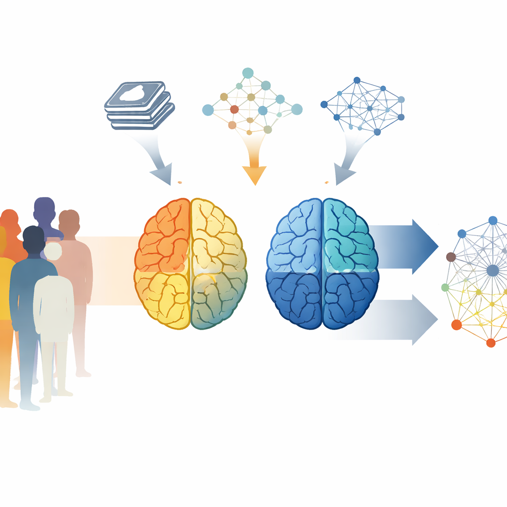
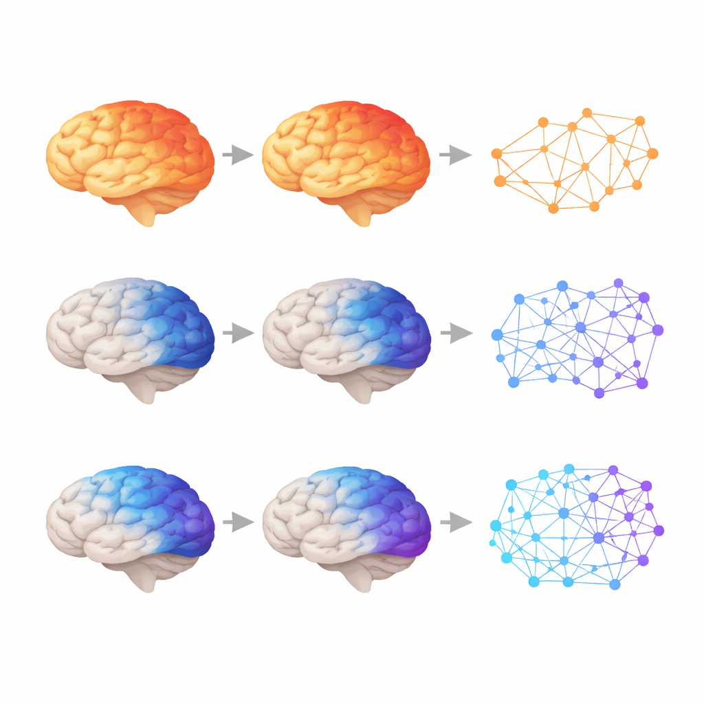
The team analysed MRI brain scans from 85 people with schizophrenia and 224 healthy volunteers. Instead of grouping all patients together, they used a data-driven tool called SuStaIn that is designed to detect hidden subtypes and stages of disease from cross-sectional data. This approach allowed them to reconstruct how brain tissue loss, or atrophy, seems to spread across the brain over the course of illness. The analysis revealed two clear subtypes. In one group, called Subtype0, changes began in the front of the brain and in emotion-related deep structures, then moved backward. In the other, Subtype1, changes started in visual and deep relay areas toward the back of the brain and then spread forward. Both paths eventually affected wide networks, but they took opposite routes to get there.
How symptoms and thinking differ
These brain-based subtypes were not just mathematical curiosities; they lined up with real-world symptoms. People following the front-first path (Subtype0) showed more intense “positive” symptoms—experiences added to normal life, such as delusions and hallucinations—and a stronger tendency toward hostility as the disease stage advanced. Those on the back-first path (Subtype1) were more prone to social withdrawal, a “negative” symptom marked by pulling away from others, even when controlling for how advanced their illness appeared. Interestingly, Subtype0 performed somewhat better on a verbal fluency test, which taps the ability to rapidly retrieve and organize words, suggesting that the two brain patterns are linked to different profiles of thinking and behavior.
Opposite patterns in brain communication
Beyond structure, the researchers examined resting-state functional MRI, which captures how strongly different brain regions synchronize their activity when a person is lying still. Here too, the two subtypes diverged. As Subtype0 progressed, a key connection between a region near the back of the brain involved in integrating information (the angular gyrus) and a gateway area important for memory (the entorhinal cortex) weakened. This “hypoconnectivity” hints that the brain’s systems for tying together inner thoughts and memories may gradually fall apart, helping to explain problems in distinguishing inner experiences from outside reality. In contrast, as Subtype1 advanced, several connections—especially those linking visual areas, emotional hubs, and deep reward and arousal centers—grew stronger. This “hyperconnectivity” may be the brain’s attempt to compensate for early damage in sensory and deep structures, but it could also distort how sights, feelings, and motivation are combined.
Making sense of decades of mixed findings
For years, brain imaging studies of schizophrenia have disagreed on whether patients show too little or too much connectivity among brain regions. This work suggests that both sides may be right, but they have been looking at mixtures of different subtypes and disease stages. If some patients are in a front-first, weakening-connections trajectory while others are in a back-first, strengthening-connections trajectory, then averaging them together would naturally produce inconsistent results. By separating patients into biologically grounded subtypes and estimating how far along each person is in their individual trajectory, this study offers a way to untangle those contradictions.
What this could mean for care
To a layperson, the main message is that schizophrenia is unlikely to be a single, uniform brain disorder. Instead, there appear to be at least two distinct routes through which the illness reshapes the brain—one marked by gradual disconnection of key thinking and self-related networks, and another by rising, possibly overactive communication in circuits linking vision, emotion, and motivation. Recognizing these different paths could eventually help clinicians match treatments to the biology of each patient, choosing therapies that either calm down overactive circuits or support weakening ones at just the right time. While the study is cross-sectional and cannot yet prove how individuals change over years, it lays an important foundation for more precise, subtype-specific approaches to understanding and treating schizophrenia.
Citation: Yoshimaru, D., Ouchi, K., Shibukawa, S. et al. Data-driven schizophrenia subtyping via brain atrophy trajectories and functional connectivity. Transl Psychiatry 16, 229 (2026). https://doi.org/10.1038/s41398-026-03968-w
Keywords: schizophrenia subtypes, brain connectivity, MRI brain imaging, precision psychiatry, brain atrophy