Clear Sky Science · en
Soluble epoxide hydrolase in the liver orchestrates abdominal aortic aneurysm formation in mice
Why this matters for heart health
Abdominal aortic aneurysms are silent bulges in the main artery of the belly that can suddenly burst and cause deadly internal bleeding. Today, doctors mostly watch and wait, offering surgery only when the aneurysm becomes large enough to justify the risks. This study reveals an unexpected partner in crime: the liver. The researchers show that a common liver enzyme helps drive the inflammation that weakens the aortic wall, and that blocking this enzyme in mice can prevent aneurysms from forming. Their findings point toward a new kind of medicine that might one day slow or stop this dangerous disease.
A hidden conversation between liver and artery
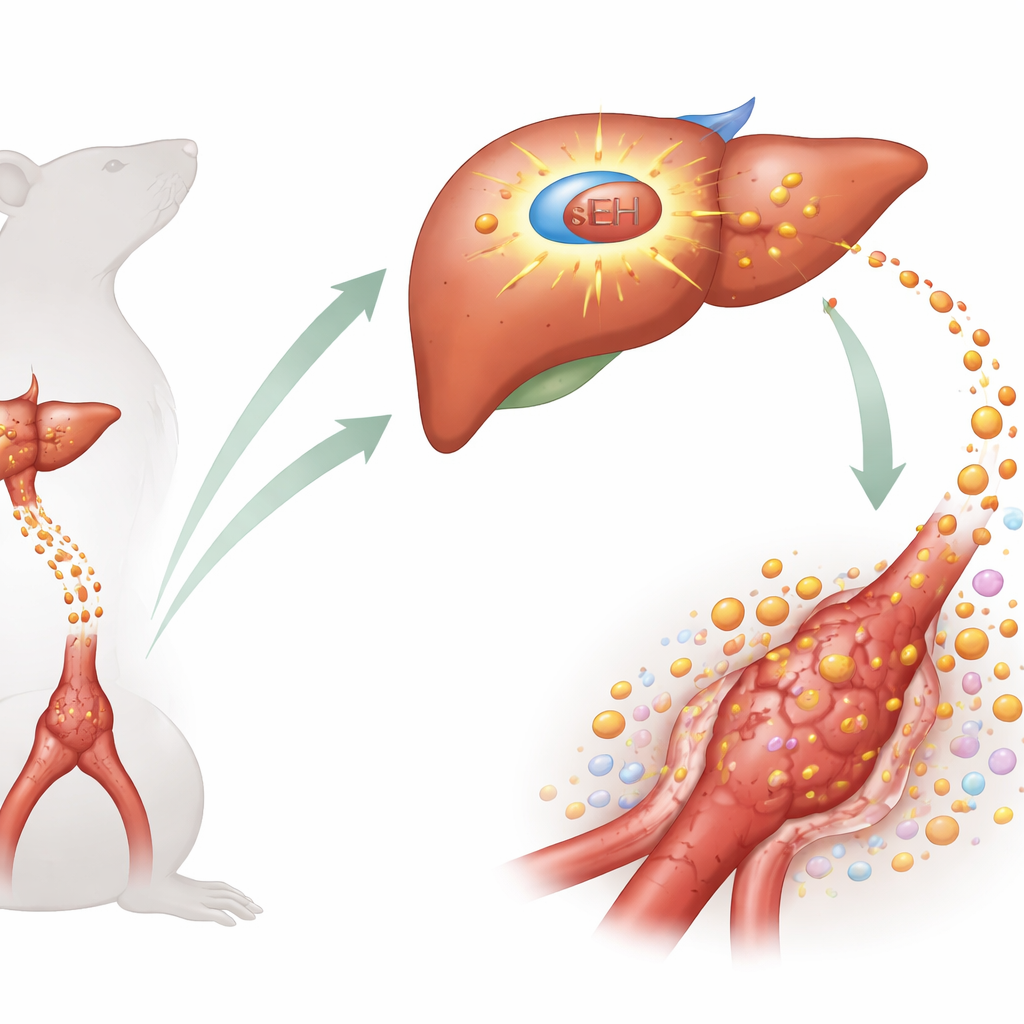
The team focused on abdominal aortic aneurysm (AAA), a condition responsible for tens of thousands of deaths worldwide each year, especially in older men and smokers. They suspected that the liver, which churns out many inflammation-related proteins, might play a bigger role in AAA than previously thought. A particular enzyme in liver cells, called soluble epoxide hydrolase (sEH), is known to turn certain fatty-acid molecules from calming, protective forms into more aggressive ones. Because sEH is far more active in the liver than in blood vessels, the authors asked whether liver-based sEH might be orchestrating the inflammation that slowly erodes the aortic wall.
When the liver turns up the inflammatory heat
Using two different mouse models of AAA—one driven by a hormone infusion and the other by a local chemical injury to the aorta—the researchers found that sEH levels and activity in the liver surged as aneurysms developed, while levels in the aorta itself stayed low. When they gave mice a highly selective drug that blocks sEH, or genetically deleted sEH only in liver cells, aneurysms were much less likely to form and existing aortic bulges were smaller. The treated mice showed fewer breaks in the elastic fibers of the aorta, less invasion by immune cells, and lower amounts of tissue-remodeling enzymes that normally help destroy the vessel wall. At the same time, the liver produced less of two powerful inflammatory proteins, complement C3 and serum amyloid A, which are known to circulate in the blood and settle in damaged arteries.
Signals from a damaged artery to the liver
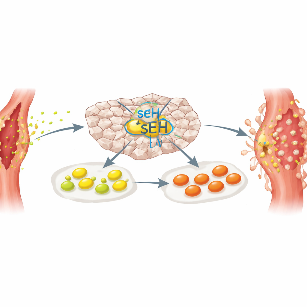
The communication was not one-way. In a clever set of experiments, the scientists grew healthy liver slices together with injured aortas taken from mice before aneurysms had fully formed. Simply placing the damaged aorta next to the liver tissue was enough to boost sEH activity and inflammatory gene expression in the liver. Protein profiling of the fluid surrounding the injured aortas pointed to several candidate messengers, and one in particular stood out: galectin-3, a sugar-binding protein released by stressed or dying cells. When the team bathed liver tissue in purified galectin-3, sEH and liver inflammatory factors rose sharply—but this effect disappeared in liver tissue lacking sEH. The results suggest that an injured aorta sends distress signals to the liver, which in turn switches on sEH and ramps up systemic inflammation.
A harmful fat messenger in the bloodstream
The researchers then searched for specific fatty-acid byproducts made by sEH that might link liver activation to aneurysm growth. They homed in on 12,13-DiHOME, a molecule derived from linoleic acid, a common component of vegetable oils and processed foods. In mice with aneurysms, levels of 12,13-DiHOME were elevated in blood and liver-derived fluids and dropped when sEH was blocked. Mice engineered to lack liver sEH had lower levels of this molecule and higher levels of its more benign precursor. When normal liver slices were exposed to 12,13-DiHOME, they secreted more complement C3 and serum amyloid A, whereas related fatty molecules had little effect. Importantly, blood samples from people with AAA also showed higher 12,13-DiHOME levels than matched individuals without aneurysms, hinting that this pathway operates in humans as well.
New paths toward gentler treatments
Taken together, the work outlines a loop in which a damaged aorta releases distress molecules, such as galectin-3, that activate sEH in the liver. The liver then floods the bloodstream with inflammatory proteins and a specific fatty-acid product, 12,13-DiHOME, which accumulate in the aortic wall and help aneurysms grow. Breaking this loop by inhibiting sEH in the liver greatly reduced aneurysm formation in mice, without lowering blood pressure or cholesterol. Several sEH-blocking drugs are already in early human trials for other conditions, raising the possibility that they could eventually be repurposed to slow AAA progression. While more research in people is needed, this study reshapes our view of aneurysms from a purely local vessel problem into a disease of long-distance cross talk between the aorta and the liver.
Citation: Kim, D.S., Horimatsu, T., Ogbi, M. et al. Soluble epoxide hydrolase in the liver orchestrates abdominal aortic aneurysm formation in mice. Commun Biol 9, 504 (2026). https://doi.org/10.1038/s42003-026-09765-x
Keywords: abdominal aortic aneurysm, liver inflammation, soluble epoxide hydrolase, fatty acid metabolites, galectin-3