Clear Sky Science · en
Fluorescence imaging for assessing tissue perfusion after revascularization in peripheral arterial disease
Why blood flow in the legs matters
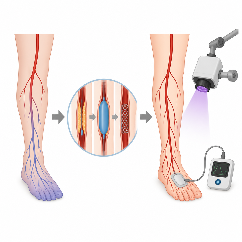
When arteries in the legs become narrowed or blocked, the muscles and skin can suffer from a lack of oxygenated blood. This condition, called peripheral artery disease, is common in older adults and people with diabetes, and it can lead to pain when walking, slow wound healing, and even risk of limb loss. Doctors have several ways to restore blood flow, but they also need reliable tools to check whether the repair has truly improved circulation in the tissues that need it most.
Current ways to check circulation
In everyday practice, doctors often rely on the ankle brachial index, which compares blood pressure in the ankle and the arm, and on measurements of skin oxygen levels using small sensors on the foot, known as transcutaneous oxygen pressure. These methods are well established, but each has drawbacks, especially in patients with diabetes or kidney disease. A newer method, fluorescence imaging with a dye called indocyanine green, can create glowing pictures of blood flow in real time. Because it looks impressive and is already used in many types of surgery, some have hoped it could become a quick, visual way to judge whether leg arteries have been successfully reopened.
A closer look at fluorescent dye imaging
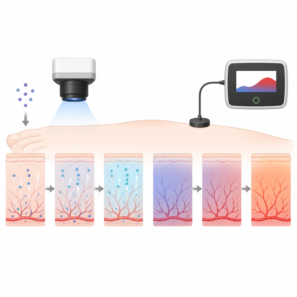
In this study, researchers followed 131 patients who underwent procedures to improve blood flow in their legs, such as balloon widening of arteries or bypass surgery. Ninety five patients with technically successful repairs and usable data were included in the final analysis. Each patient had three tests before and shortly after treatment: the ankle brachial index, skin oxygen measurement on the top of the foot, and fluorescence imaging after injection of the dye. The team focused on a small square of skin between two bones of the foot and used custom software to turn the glow of the dye over time into three numbers that describe how quickly and how strongly blood arrives.
What the fluorescent images showed
Based on how these three dye related numbers changed, the researchers grouped patients into three categories: those whose local perfusion seemed improved, those whose perfusion looked unchanged, and those whose perfusion appeared worse after the operation. About two thirds of patients fell into the improvement group, while nearly one in five seemed to show worse perfusion according to the dye curves. In the improvement group the brightness rose faster and higher and reached its peak sooner, while in the worsened group it rose more slowly, to a lower peak, and peaked later. On the surface this suggested that fluorescence imaging can clearly register changes in blood flow at the foot.
How the new method compared with trusted measures
The crucial test was whether these changes in the dye signal matched other indicators. The ankle brachial index and skin oxygen levels both improved significantly after the procedures, as expected when blocked arteries are successfully treated. Skin oxygen values also tracked with how much patients improved in symptom scores and wound healing over the next three months. In contrast, the dye based measurements did not line up with either the ankle pressure test, the skin oxygen readings, or the patients’ clinical progress. Some patients whose fluorescence imaging looked worse actually had better ankle pressures, higher skin oxygen, and clear symptom relief.
What this means for patients and doctors
For patients, the key message is that eye catching glowing images are not necessarily the best guide to how well blood is reaching at risk tissues in the foot. In this large, carefully monitored group, standard skin oxygen measurements remained a more dependable indicator of improvement after artery repair than dye based fluorescence imaging. Because the dye requires a vein injection and cannot be used safely in all patients, and because its measurements did not match established tests or outcomes, the authors conclude that fluorescence imaging should not replace traditional methods for checking tissue perfusion after leg revascularization.
Citation: Kluckner, M., von Kroge, P.H., Duprée, A. et al. Fluorescence imaging for assessing tissue perfusion after revascularization in peripheral arterial disease. Sci Rep 16, 15967 (2026). https://doi.org/10.1038/s41598-026-47505-7
Keywords: peripheral artery disease, tissue perfusion, fluorescence imaging, transcutaneous oxygen pressure, revascularization