Clear Sky Science · en
Predicting the degree of trait emotional empathy from cortical features using surface-based morphometry
Why caring about caring matters
Most of us know people who seem to deeply feel what others are going through, and others who find that harder. This study asks whether such differences in emotional empathy are reflected in the physical structure of the brain. By combining brain scans with a questionnaire about everyday feelings for others, the researchers explored how subtle features on the brain’s surface relate to how strongly people tend to share other people’s emotions.
Looking for empathy in the brain
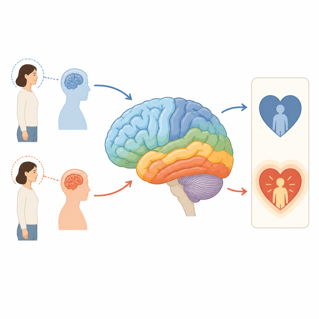
The team focused on emotional empathy, the tendency to actually feel echoes of another person’s joy or pain, rather than just understanding it in a detached way. Sixty two adults in the Czech Republic completed the Toronto Empathy Questionnaire, which measures how often they experience such emotional reactions in daily life. They also underwent high resolution MRI brain scans. Instead of using older methods that look at chunky blocks of brain tissue, the researchers used a more refined approach that traces the brain’s outer surface and measures how thick the cortex is, how folded it is, and how deep the grooves run.
Key spots on the brain’s surface
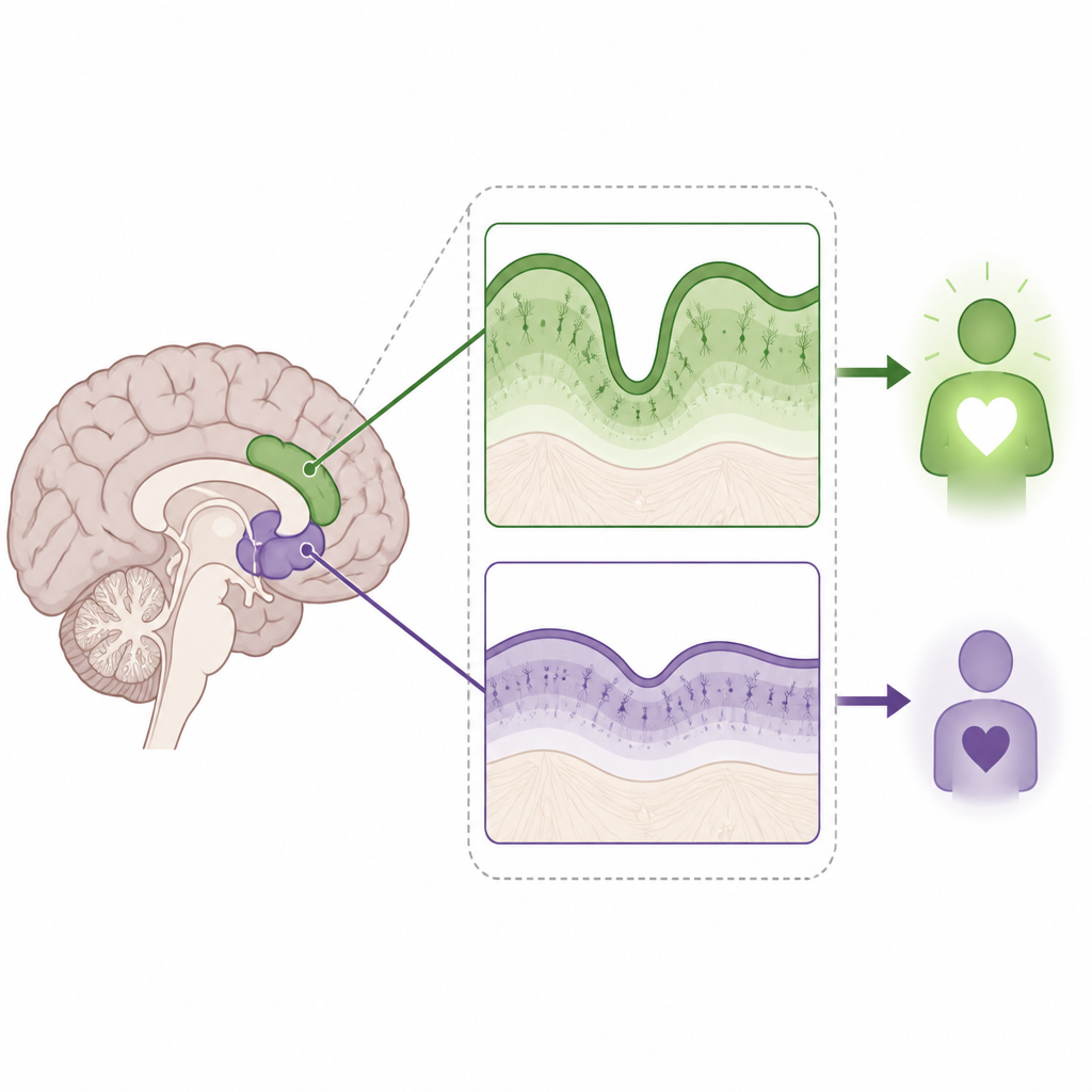
Previous brain imaging work has shown that certain regions light up when people are actively feeling with someone else during an experiment. Two such regions are the insula, tucked deep within the side of the brain, and a strip of tissue toward the front called the dorsal anterior cingulate cortex. The researchers reasoned that if momentary, in the moment empathy and long term, trait empathy rely on the same systems, then the physical properties of these regions might track with a person’s general level of emotional empathy. They tested this idea by examining how the thickness of these areas related to questionnaire scores, while also considering age and gender.
Thinner in some regions, higher in empathy
Surprisingly, the results ran in the opposite direction of what the team had predicted. People with higher emotional empathy tended to have a thinner cortex in the left insula and in the left dorsal anterior cingulate, not a thicker one. The right insula did not show a clear link with empathy at all. When the researchers allowed for more complex patterns rather than a simple straight line relationship, they found that the left insula showed a curved pattern: within a certain range, changes in thickness related to empathy in a non linear way. This hints that there may be an optimal balance of tissue in that region for supporting emotional resonance with others.
Why brain shape could not forecast empathy
Beyond looking at these few regions, the scientists also tried to predict each person’s empathy score from detailed measurements across hundreds of small patches covering the whole cortex. They used a machine learning method designed to sift through many correlated features and pick out the most informative ones. On the original data, some models seemed to fit reasonably well, especially for how folded the surface was. But when the models were tested on new data using leave one out cross validation, their performance dropped sharply. In fact, they did worse than simply guessing the average empathy score for everyone, suggesting that, in this modest sample, brain shape alone could not reliably forecast how empathic a new person would be.
What this means for understanding empathy
The study suggests that people who more strongly feel others’ emotions tend to have slightly thinner tissue in certain emotion related brain regions, and that this relationship may not be purely linear. One possible explanation is that over development, fine tuning processes may trim back excess connections, leaving a more efficient but thinner cortex that supports rich emotional responses. At the same time, the failed prediction tests are a reminder that reading someone’s empathy level from a brain scan is far from ready for real world use. Emotional empathy appears to arise from subtle, distributed features that are hard to capture with structure alone and in small groups of volunteers.
Looking ahead
For a lay reader, the main takeaway is that our capacity to feel with others does have detectable links to the physical shape of specific brain areas, but these links are modest and complex. Brain scans in this study could not yet predict who is more empathic on an individual basis, mainly because the dataset was small compared with the huge number of brain features measured. Future work with larger groups and additional measures, such as the wiring of white matter pathways, may paint a more complete picture of how the brain supports the everyday human ability to share in one another’s feelings.
Citation: Novak, L., Malinakova, K., van Dijk, J.P. et al. Predicting the degree of trait emotional empathy from cortical features using surface-based morphometry. Sci Rep 16, 14893 (2026). https://doi.org/10.1038/s41598-026-44137-9
Keywords: emotional empathy, brain structure, cortical thickness, MRI study, social neuroscience