Clear Sky Science · en
Body fat percentage and sacral–abdominal wall distance are associated with obesity in patients with osteoporosis: a retrospective study
Why this study matters for everyday health
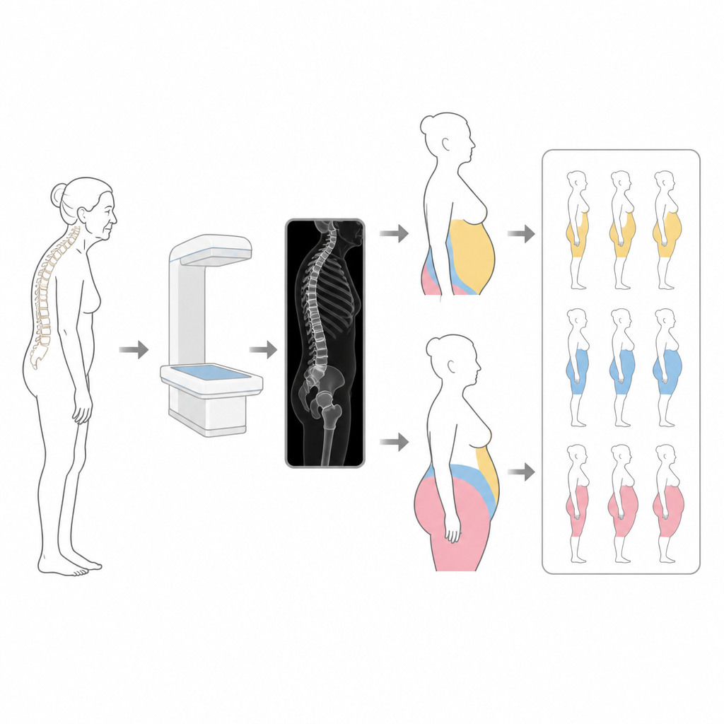
As people live longer, more older adults are living with both fragile bones and extra body fat. Doctors usually rely on body mass index, or BMI, to judge whether someone has obesity. But in people with osteoporosis, curved spines and height loss can make BMI misleading. This study explores two simple measurements that might give a clearer picture of body fat in older women with weak bones and bent backs.
Looking beyond the bathroom scale
The researchers focused on 385 Japanese women aged 65 and older who were being treated for osteoporosis. Instead of judging weight status by BMI alone, they also used body fat percentage from a full-body scan and a new X-ray based measure of belly shape. Their goal was to see whether these extra measurements could better identify who was truly carrying risky amounts of fat, even if their BMI looked normal.
Two ways to describe belly fat
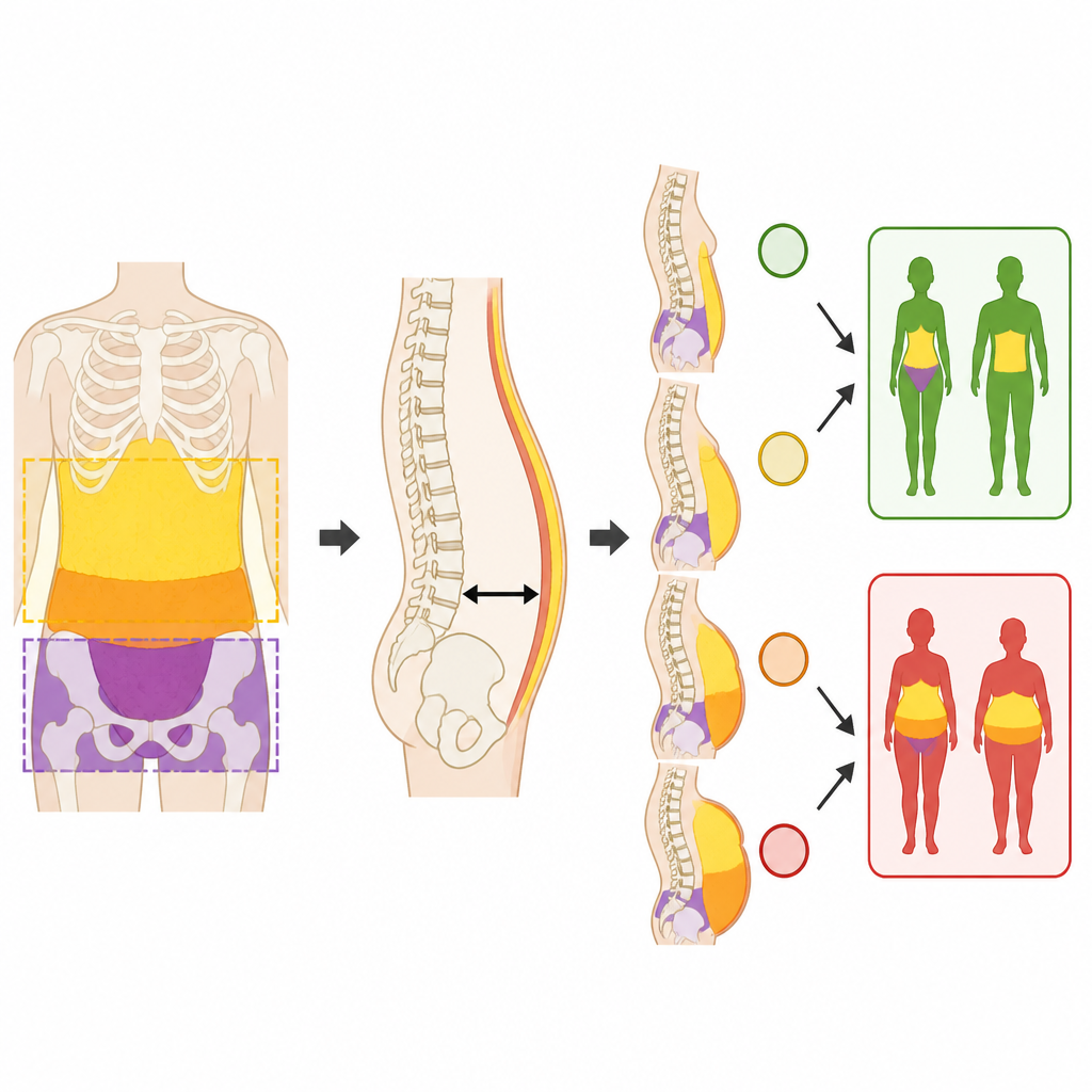
One key measure was the android to gynoid fat ratio, which compares fat stored around the waist to fat around the hips and thighs. A higher ratio means more upper-body fat and is thought to be more harmful for the heart and metabolism. The second measure was the sacral–abdominal wall distance, taken from a side-view spinal X-ray while standing. This distance reflects how far the belly sticks out in relation to the spine and pelvis, capturing not just fat but also posture and spinal curve.
What the study found
The team defined obesity in two ways: first, by the usual Japanese BMI cut-off of 25 or higher, and second, by either that BMI cut-off or a body fat percentage of at least 35 percent. In both cases, women with more belly-centered fat and a larger sacral–abdominal wall distance were much more likely to be classified as having obesity. Even after taking age, height, and basic nutritional status into account, both measurements stayed strongly linked to obesity. Women who met cut-offs for both a high waist-to-hip fat pattern and a large belly distance had the highest chances of being in the obesity group.
Working together for a clearer picture
Importantly, combining the two measures told doctors more than either one alone. The fat ratio revealed where fat was stored, while the X-ray distance captured how spinal curve and posture shaped the abdomen. Together, they helped uncover women who might look only mildly overweight by BMI but actually carried a high share of fat around the waist. This approach may be especially helpful for patients whose height has shrunk because of spinal fractures, which can make BMI seem reassuring even when unhealthy fat is present.
What this means for patients and clinicians
The study suggests that in older women with osteoporosis, checking both body fat distribution and belly shape may give a truer picture of obesity than BMI alone. However, the research looked only at one point in time, so it cannot show whether these measures actually cause health problems or predict future illness. Still, the findings hint that simple scans and X-rays, which many osteoporosis patients already receive, could be used in new ways to guide conversations about weight, body shape, and overall health.
Citation: Nagai, T., Kasai, F., Sugiyama, M. et al. Body fat percentage and sacral–abdominal wall distance are associated with obesity in patients with osteoporosis: a retrospective study. Sci Rep 16, 15669 (2026). https://doi.org/10.1038/s41598-026-43802-3
Keywords: osteoporosis, obesity, body fat, spinal curvature, older women