Clear Sky Science · en
Pro-tumoral Ca2+ signaling is dependent on Slowpoke and Ca-α1T channels in Drosophila melanogaster glioma
Why Brain Tumors Talk to the Nervous System
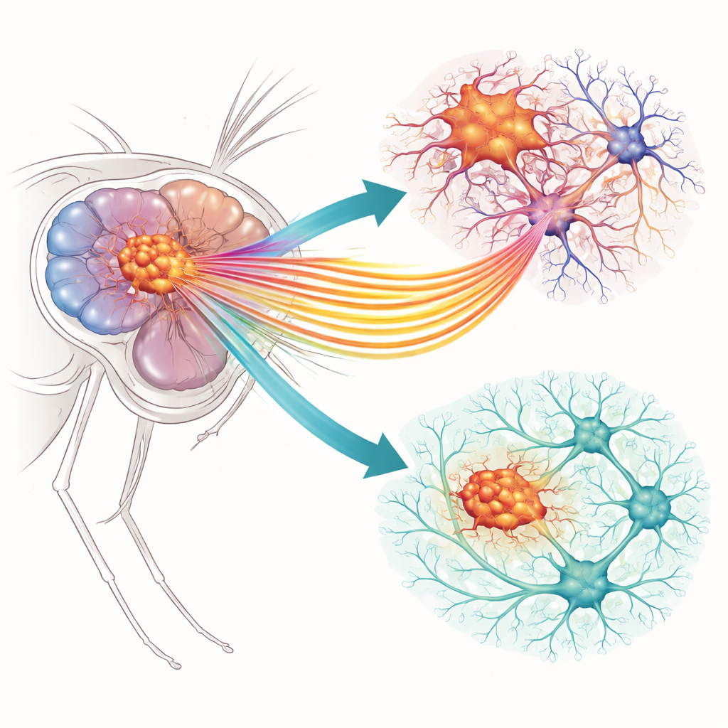
Glioblastoma, a deadly form of brain cancer, doesn’t grow in isolation. It weaves itself into the brain’s own wiring, hijacking normal signals to fuel its spread and damage nearby neurons. This study uses the fruit fly, Drosophila melanogaster, to uncover how tiny pores in cell membranes, called ion channels, help tumor cells amplify internal calcium signals that drive growth, reshape their connections, and ultimately shorten lifespan. By pinpointing which channels matter most, the work suggests new ways to slow aggressive brain tumors in more complex animals, including humans.
Two Tiny Gates with Big Consequences
The researchers focused on two specific ion channels in glial tumor cells, the brain’s support cells gone rogue. One, called Slo, lets potassium ions out of the cell when calcium levels rise. The other, Ca-α1T, is a calcium channel that lets calcium ions in when the cell membrane is activated. Their close mammalian counterparts have already been linked to human glioblastoma in cell cultures, but their behavior inside a living brain network was less clear. Using a well-established fly model in which glial cells carry cancer-like mutations in growth pathways (EGFR and PI3K), the team could compare normal brains with tumor-bearing ones and ask how dialing down each channel affected tumor behavior and the surrounding nervous system.
Turning Down the Volume on Tumor Growth
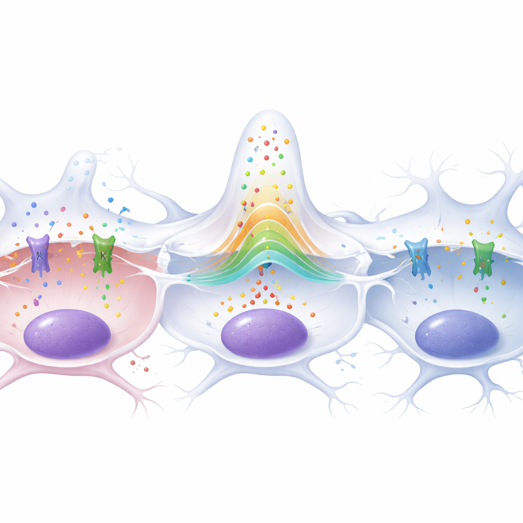
First, the scientists confirmed that both channels are present in glial cells of healthy and tumor-bearing fly brains. Ca-α1T showed somewhat higher levels in tumor cells, while Slo levels were similar in normal and glioma glia. They then used targeted genetic tools (RNA interference) to reduce the amount of each channel only in glial cells. In flies with gliomas, lowering either Slo or Ca-α1T sharply reduced the number of tumor glial cells and shrank the swollen glial membrane network that normally wraps deep into brain tissue. Measurements of a calcium-activity reporter revealed that glioma cells normally show strikingly elevated calcium signaling, but this hyperactivity dropped back to near-normal when either channel was silenced. At the same time, markers of two major growth-driving pathways, ERK and PI3K, were also reduced, showing that these membrane channels help feed the tumor’s internal growth circuitry.
Protecting Neurons and Extending Life in Flies
Although both channels helped drive proliferation, they did not have identical effects on the brain. The team counted synapses at the neuromuscular junction, a sensitive readout of neuron health in the fly. Glioma-bearing larvae lost about 70 percent of these synapses, reflecting strong tumor-induced neurodegeneration. Reducing Ca-α1T did not rescue this loss. In contrast, cutting down Slo in tumor glia significantly restored synapse numbers toward normal, indicating that Slo has a special role in how glioma cells harm neurons. This difference became even clearer in adult flies: animals with gliomas died much earlier than healthy controls, but when Slo was reduced in the tumor cells, their survival curves nearly overlapped with non-tumor flies. Lowering Ca-α1T, by comparison, did not extend lifespan and even shortened survival in healthy flies.
Rewiring Metabolism and Chemical Communication
To see how these channels reshape tumor cell behavior at the gene level, the authors performed RNA sequencing on fly heads. Tumor brains showed broad shifts in metabolism and synaptic signaling, including increased activity in carbohydrate use and glutamate-based communication, both hallmarks of aggressive gliomas. Turning down Ca-α1T mainly dampened genes involved in sugar breakdown and storage, including enzymes tied to the “Warburg effect,” where cancer cells rely heavily on altered glucose metabolism. Reducing Slo, on the other hand, selectively lowered genes encoding key glutamate receptor subunits and cell-adhesion molecules that help glioma cells form and stabilize connections with neurons. One of these, similar to human AMPA-type glutamate receptors, has previously been linked to tumor overgrowth and poor outcomes, while another mirrors a human synaptic protein associated with worse survival when overactive.
What This Means for Future Brain Cancer Therapies
Overall, the study shows that both Slo and Ca-α1T channels act as amplifiers of calcium-dependent growth signals in glioma cells, helping them multiply, extend long membrane tubes, and keep powerful growth pathways switched on. Yet only Slo emerges as a dual lever that not only slows tumor expansion but also protects neurons and extends the lives of tumor-bearing animals. By tying Slo to abnormal synaptic communication and glutamate signaling, the work points to this channel’s human relatives as especially promising targets: blocking them might weaken the tumor, cut its harmful dialogue with neurons, and improve brain function. While fruit flies are far from patients, their shared molecular machinery makes them a valuable guide to which channels and pathways deserve the closest look in future glioblastoma therapies.
Citation: Alza, L., Montes-Labrador, P., Megías, D. et al. Pro-tumoral Ca2+ signaling is dependent on Slowpoke and Ca-α1T channels in Drosophila melanogaster glioma. Sci Rep 16, 12297 (2026). https://doi.org/10.1038/s41598-026-42712-8
Keywords: glioblastoma, calcium signaling, ion channels, glutamate receptors, brain tumors