Clear Sky Science · en
Ultra-performance liquid chromatography–mass spectrometry analysis of post-mortem brain tissue reveals specific amino acid profile dysregulation in Parkinson’s disease and Alzheimer’s disease patients
Why tiny brain chemicals matter
Parkinson’s and Alzheimer’s diseases are usually described in terms of dying nerve cells and clumps of misfolded proteins. But behind these visible changes lies a subtle chemical world of small molecules that fuel and fine‑tune brain activity. This study asks a simple but important question: do the patterns of these small building blocks, especially amino acids, change inside the brain itself in Parkinson’s disease, and are those changes different from what happens in Alzheimer’s disease and in the rest of the body? The answer could help explain symptoms, guide drug development, and point to more precise diagnostic tests.
Looking inside brains after death
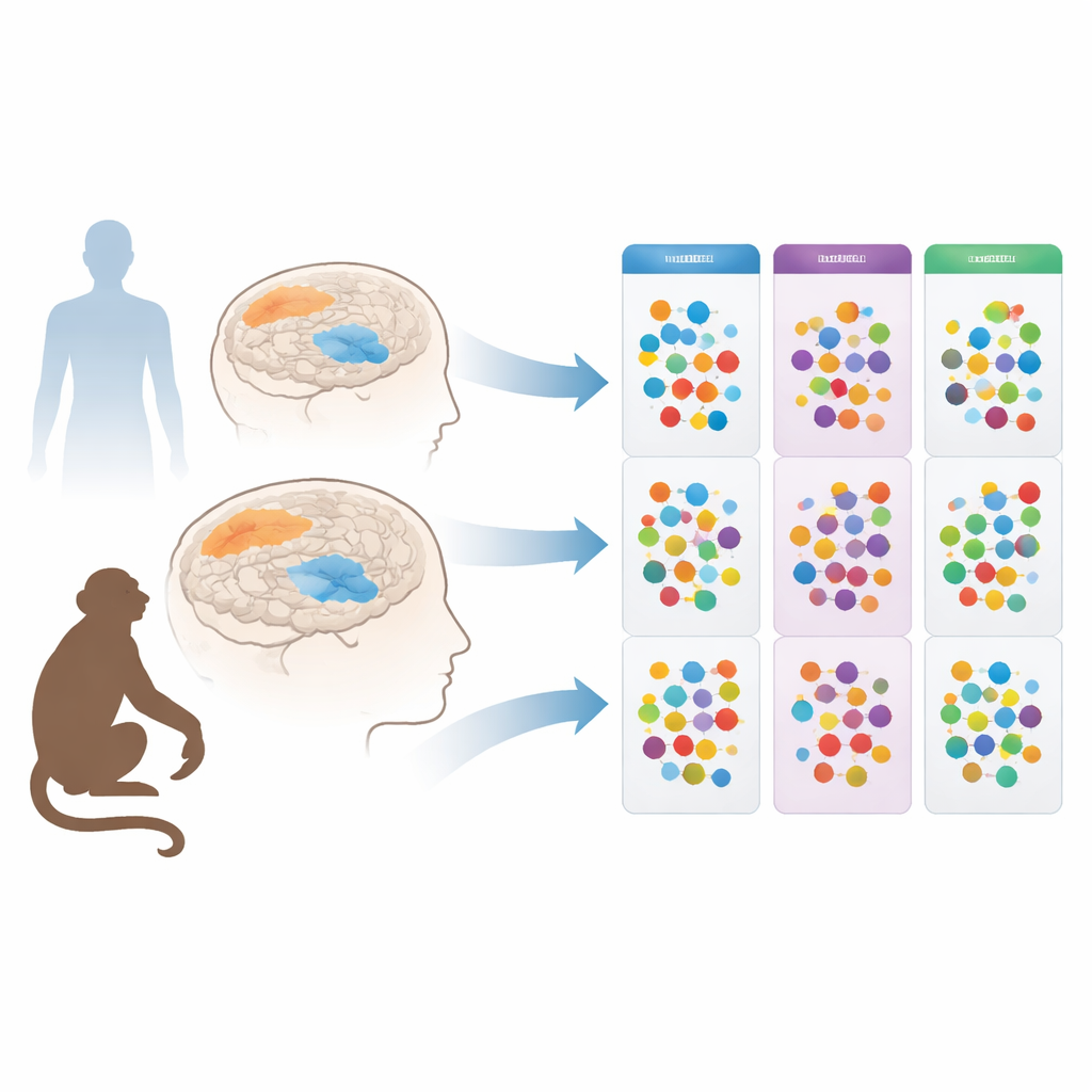
The researchers focused on amino acids, the tiny units that make up proteins and also act as messengers and energy helpers in the brain. Earlier work had shown that people with Parkinson’s have altered amino acid levels in blood, spinal fluid, saliva, and urine. However, it was unclear whether these shifts directly reflected what was happening in the brain, or were merely echoes of broader changes in organs such as the liver and kidneys. To get a clearer picture, the team turned to post‑mortem samples: they examined two brain regions in people who had died with Parkinson’s or Alzheimer’s, and in carefully matched control donors without neurodegenerative disease. They also studied an established monkey model of Parkinson’s, in which a toxin selectively damages dopamine‑producing cells, with or without long‑term treatment with the standard drug L‑DOPA.
Two brain regions, very different stories
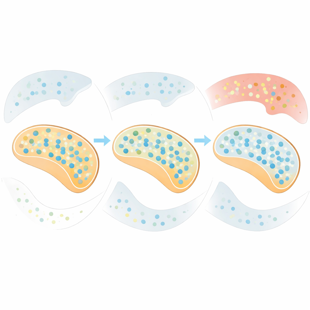
The scientists compared a deep movement‑related structure—the caudate‑putamen (or putamen in monkeys)—with a frontal thinking region called the superior frontal gyrus. Using an ultra‑sensitive technique (UPLC‑MS) that can precisely measure dozens of amino acids at once, they discovered that changes in Parkinson’s were strikingly specific to the deep movement area. In toxin‑treated monkeys, the putamen showed higher levels of several amino acids, including glutamate and aspartate (important for excitation), GABA (inhibition), the branched‑chain amino acids, phenylalanine, and serine. Adding L‑DOPA shifted the pattern further, boosting other molecules such as glycine, threonine, and citrulline. Yet in the frontal cortex of these same animals, amino acid levels were essentially unchanged, even after months of dopamine loss and treatment.
Parkinson’s versus Alzheimer’s in the human brain
Human samples told a complementary story. In the caudate‑putamen of Parkinson’s patients, only a small subset of amino acids was consistently altered. Across all stages of disease spread (Braak Lewy body stages), levels of serine were increased; in the most advanced stage, proline was also higher, while phosphoethanolamine was lower, and arginine tended to drop compared with earlier stages. These shifts suggest that as Parkinson’s progresses, certain chemical pathways—linked to energy use, antioxidant defenses, and signaling—become increasingly unbalanced. Importantly, the superior frontal gyrus of Parkinson’s patients looked essentially normal in this respect, mirroring the monkey findings and underscoring that the metabolic disruption is concentrated in the movement circuitry. In contrast, people with Alzheimer’s disease showed the opposite pattern in the same frontal region: several amino acids, including tryptophan, phenylalanine, threonine, tyrosine, and methionine, were clearly elevated, pointing to a cortical‑centered disturbance distinct from that seen in Parkinson’s.
The special role of one amino acid
Among all the molecules examined across animals, brain regions, and disease stages, serine stood out. It was consistently higher in the damaged movement area of toxin‑treated monkeys and of people with Parkinson’s, and earlier work by the same group has found elevated serine in the spinal fluid and blood of patients as well. Serine comes in two mirror‑image forms that together support both brain signaling and cellular housekeeping: one form helps activate a key glutamate receptor involved in learning and communication between neurons, while the other feeds into the production of membrane components, nucleotides, and antioxidants. The repeated rise of serine across experiments hints that the brain may be trying to adapt to dopamine loss by strengthening certain circuits and protective pathways, even as the disease advances.
What this means for patients and future tests
Viewed together, the results show that changes in small brain chemicals are not random, but follow clear patterns tied to both brain region and disease type. In Parkinson’s, amino acid shifts cluster in the dopamine‑dependent movement circuits and are relatively limited in scope, with serine emerging as a reliable marker of disturbance; in Alzheimer’s, amino acid changes are more prominent in the frontal cortex. This suggests that blood‑based metabolic fingerprints partly mirror, but do not fully capture, what happens in the brain. For a lay observer, the key takeaway is that tracking specific amino acids—especially serine—could one day help refine diagnosis, monitor how Parkinson’s is progressing, and evaluate whether new treatments are restoring healthier brain chemistry, all while distinguishing Parkinson’s from other forms of dementia.
Citation: Gervasoni, J., Di Maio, A., Serra, M. et al. Ultra-performance liquid chromatography–mass spectrometry analysis of post-mortem brain tissue reveals specific amino acid profile dysregulation in Parkinson’s disease and Alzheimer’s disease patients. npj Parkinsons Dis. 12, 95 (2026). https://doi.org/10.1038/s41531-026-01306-x
Keywords: Parkinson’s disease, amino acids, serine, brain metabolism, Alzheimer’s disease