Clear Sky Science · en
1H R1ρ relaxation identifies a hidden intermediate in DNA base-pairing
Why hidden DNA motions matter
DNA is often drawn as a tidy double helix with neatly matched base pairs, but in real life it is constantly breathing and flickering between slightly different shapes. Those brief, rare shapes can influence how DNA is repaired, read, or targeted by drugs. This study develops a more reliable way to watch some of these fast shape changes at the atomic level and, using that method, uncovers a previously hidden intermediate state in one of DNA’s most important base-pair switches—showing that even a classic anticancer drug can reshape DNA’s internal motion.
A closer look at DNA’s quiet shape changes
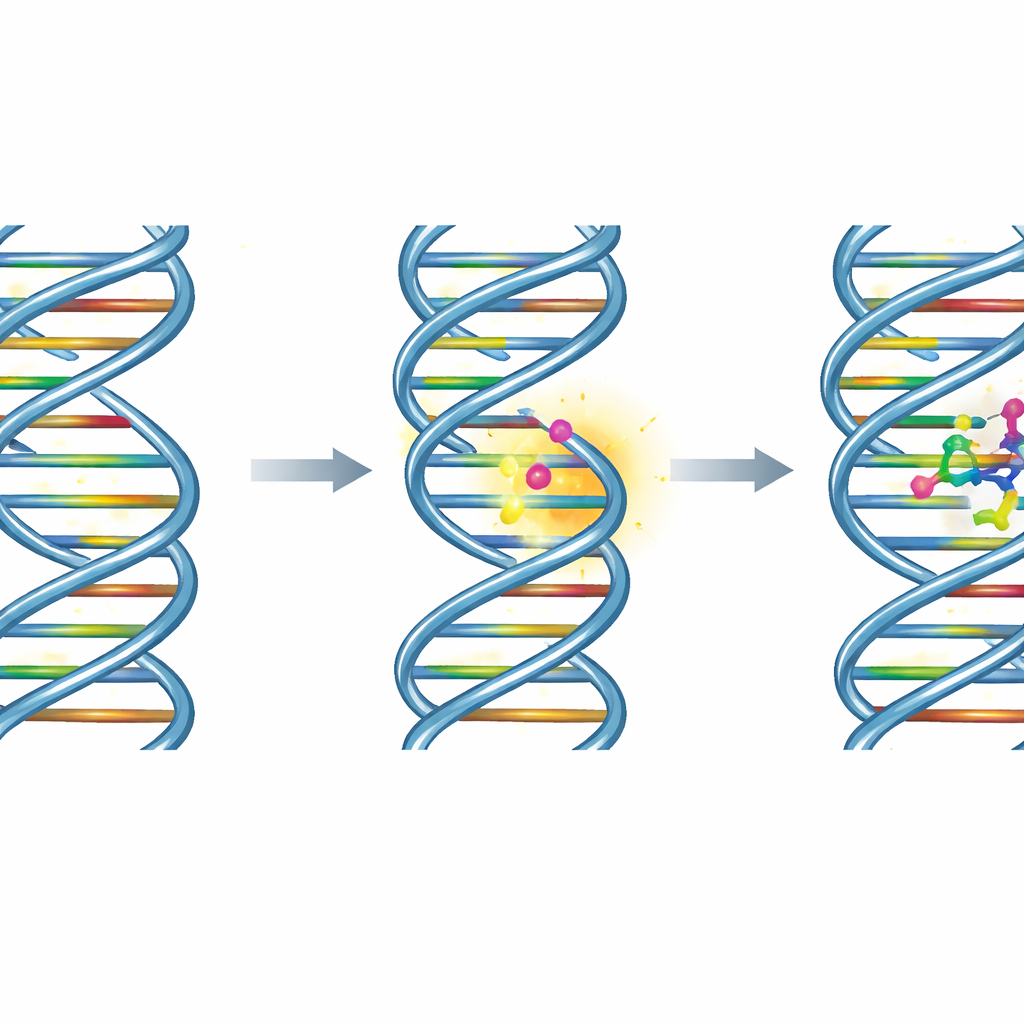
Inside every cell, the familiar A–T and G–C base pairs in DNA do not stay frozen. On time scales of millionths to thousandths of a second, they can visit rare, higher-energy arrangements. One well-known alternative is the Hoogsteen pairing, where one base in the pair briefly flips its orientation. Although such states are typically less than 2% of the population, they are linked to processes such as DNA damage repair, misincorporation of bases, and the way certain proteins recognize their targets. Because these fleeting states are so short-lived and scarce, they are nearly invisible to standard structural tools like X‑ray crystallography or cryo‑electron microscopy, but can be probed by nuclear magnetic resonance (NMR) methods that are sensitive to motion.
Sharpening an NMR tool to see the unseen
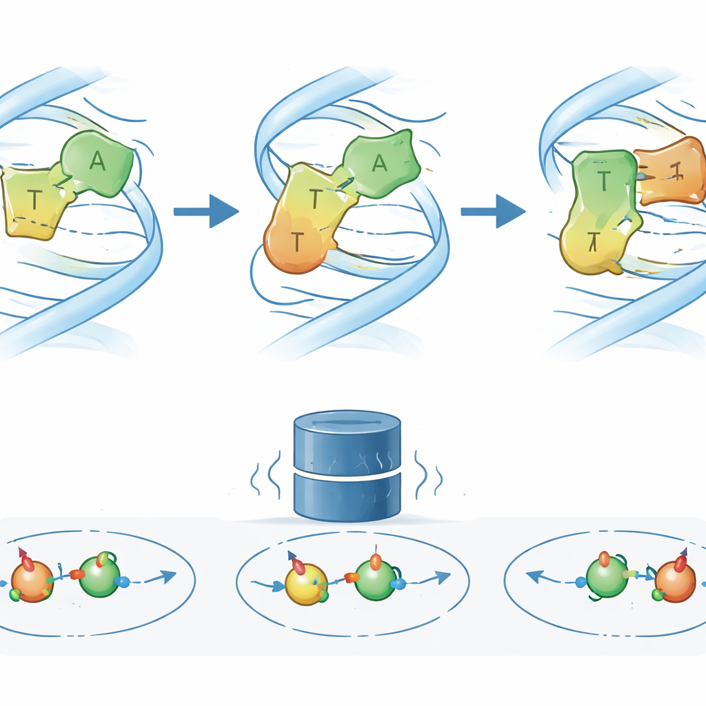
The authors focus on a specialized NMR approach called proton R1ρ relaxation dispersion, which tracks how hydrogen nuclei in DNA relax under a steady radiofrequency “spinlock” field. Changes in the relaxation rate as the field is varied encode how fast and how often the molecule jumps between different shapes. A longstanding concern, however, has been that nearby protons can “talk” to each other through cross-relaxation, creating artifacts that mimic real conformational exchange. Using detailed simulations based on established NMR equations, the team shows that for typical proton–proton separations found between stacked base pairs in DNA and RNA (around 3.4–3.9 Å), these unwanted effects are very small—within about 5% of the signal. Only when protons are closer than about 3 Å, such as within a single base pair, do artifacts become significant, and the study lays out practical rules for choosing experimental settings that avoid these pitfalls.
A hidden intermediate in DNA base pairing
Armed with this cleaner picture of what their NMR signals mean, the researchers revisit a model DNA sequence where the standard Watson–Crick–Franklin A–T pair is known to transiently convert to a Hoogsteen form. Earlier work using carbon and nitrogen NMR suggested a simple two-state switch. Proton R1ρ measurements now reveal something more complex: a third, low-population excited state (ES2) that sits between the usual and Hoogsteen arrangements. By tracking the chemical shifts and exchange rates of several hydrogen atoms in and around the key base pair, the team finds that ES2 is a consistent feature of neighboring A–T pairs and connects to both the ground state and the Hoogsteen state, forming a three-state network rather than a simple on–off toggle.
Probing the new state with designer bases and a cancer drug
To understand what ES2 might look like, the authors chemically altered one partner base in the key A–T pair, removing or changing specific atoms involved in hydrogen bonding. These “designer” bases selectively weaken or favor different pairing modes. The resulting changes in NMR signals and exchange rates show that when a particular amino group is removed, the DNA samples ES2 much more often, implying that this group normally stabilizes the ground-state pairing and slows access to the intermediate. Advanced molecular simulations, combined with quantum-based predictions of NMR shifts, then point to a plausible structural model: in ES2, the adenine base partially reorients so that its amino group forms a temporary hydrogen bond to an alternative oxygen atom on thymine, loosening the original bond network on the way toward a Hoogsteen-like geometry. Strikingly, when the anticancer drug actinomycin D is added, the NMR “fingerprint” of ES2 becomes more prominent, indicating that the drug stabilizes or enriches this intermediate as it binds along the DNA.
What this means for DNA biology and drug action
By quantifying when and how proton-based R1ρ measurements can be trusted, this work turns a specialized NMR experiment into a more broadly usable tool for watching fast, rare motions in DNA and RNA. Using that improved lens, the authors uncover a drug-stabilized, previously hidden intermediate in the classic A–T base-pair switch between normal and Hoogsteen forms. For non-specialists, the key message is that DNA is not just a static code but a shifting landscape of states, and some medicines may work in part by reshaping that landscape—favoring particular fleeting structures that influence how DNA is read, damaged, or repaired.
Citation: Dasgupta, R., Steinmetzger, C., Ilgen, J. et al. 1H R1ρ relaxation identifies a hidden intermediate in DNA base-pairing. Nat Commun 17, 4114 (2026). https://doi.org/10.1038/s41467-026-72559-6
Keywords: DNA base-pair dynamics, NMR relaxation dispersion, Hoogsteen base pairs, conformational intermediates, actinomycin D