Clear Sky Science · en
Structures of respiratory supercomplexes and ATP synthase oligomers in mammalian mitochondrial inner membrane
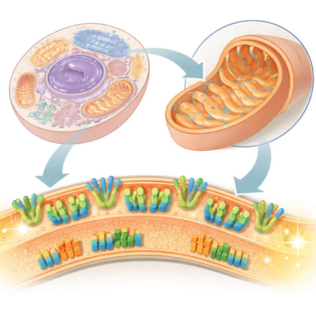
Power Plants Inside Our Cells
Every second, trillions of tiny machines inside your cells churn out the energy that keeps you alive. This paper takes an unusually close look at some of the most important of these machines inside mitochondria, the so‑called “power plants” of the cell. Using cutting‑edge electron microscopy, the authors reveal how energy‑producing protein complexes sit in their natural membrane environment, how they team up into larger assemblies, and how their shapes help sculpt the inner architecture of mitochondria itself. These details matter because subtle defects in these structures are linked to metabolic diseases and mitochondrial disorders.
The Inner Landscape of a Power Plant
Mitochondria have two membranes, and the inner one is where the real energy business happens. It hosts two major sets of protein machines: the respiratory chain, which moves electrons and pumps protons to create a voltage across the membrane, and ATP synthase, which uses that voltage to make ATP, the cell’s “energy currency.” Traditionally, scientists have studied these proteins after extracting them with detergents, which risks disturbing fragile attachments and lipids. In this work, the researchers use sub‑mitochondrial particles—small vesicles peeled from bovine heart mitochondria—and image them directly in a frozen, native‑like state using cryo‑electron microscopy. This approach lets them see not just individual proteins, but how those proteins are arranged and cooperate in the actual membrane.
Shaping the Ridges That Make Energy
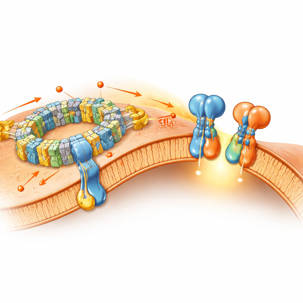
One of the most striking findings concerns ATP synthase, the rotary enzyme that manufactures ATP. Previous studies suggested that two ATP synthase units can pair to form a V‑shape, and that larger groups might bend the inner membrane into tight ridges called cristae, but it was unclear whether the larger assemblies were natural or artifacts from the extraction process. Here, the authors clearly see ATP synthase dimers joined by a small regulatory protein, forming pairs that are embedded in the membrane. Even more importantly, they observe linear tetramers—two dimers arranged side by side—right in the native membrane. These tetramers sit at sharply curved regions and collectively bend the membrane into a U‑shaped profile, supporting the idea that ATP synthase assemblies actively shape cristae tips in mammalian mitochondria.
Fine Details of the Rotary Machine
The study also zooms in on the membrane‑spanning portion of ATP synthase, uncovering details that challenge earlier interpretations. A ring of protein subunits (the c8‑ring) rotates within the membrane, and earlier detergent‑based structures showed extra density inside this ring, thought to be tightly held lipids interacting with another subunit (called e). In the native membrane, however, the authors find that this inner density is very weak or absent, suggesting that what was previously seen may actually have been detergent molecules, not essential lipids. Instead, their maps hint that the end of the e‑subunit, possibly bearing a small chemical modification, interacts directly with the ring. This subtle rearrangement changes how scientists imagine the mechanical linkage that allows the membrane’s voltage to drive ATP production.
Energy Machines Team Up
Beyond ATP synthase, the paper explores how the respiratory chain complexes—numbered I, III, and IV—cluster into “supercomplexes.” In their native membrane samples, the authors find not only the previously known combinations (such as one complex I with a dimer of complex III and one or two copies of complex IV), but also a new form containing three copies of complex IV attached to the core unit, and even a giant “megacomplex” that holds two complex I units, a dimer of complex III, and six copies of complex IV. These higher‑order assemblies curve the membrane slightly and likely optimize how electrons and protons move, making energy conversion more efficient. At the same time, the individual complexes largely retain the same fine‑scale structures seen in traditional purified samples, indicating that many of their core features survive detergent‑based preparation.
Implications for Health and Disease
By preserving the natural setting of these protein machines, this work provides a more faithful snapshot of how mitochondrial “hardware” is organized in living cells. The authors show that ATP synthase tetramers are genuine features of mammalian mitochondria and that they help mold the sharp ridges where ATP production is concentrated. They also reveal a richer variety of respiratory supercomplexes and megacomplexes than previously recognized. Because mutations in these complexes and changes in how they assemble are linked to metabolic disorders, mitochondrial diseases, and even the early steps of cell death, this structural map offers a solid foundation for future studies. In simple terms, the paper explains how the layout and teamwork of the cell’s tiniest turbines and wires help keep our energy supply running smoothly—and how subtle mis‑wiring might contribute to human disease.
Citation: Nakano, A., Masuya, T., Akisada, S. et al. Structures of respiratory supercomplexes and ATP synthase oligomers in mammalian mitochondrial inner membrane. Nat Commun 17, 4075 (2026). https://doi.org/10.1038/s41467-026-70578-x
Keywords: mitochondria, ATP synthase, respiratory supercomplexes, cryo electron microscopy, mitochondrial diseases