Clear Sky Science · en
Identification of a new population of Tnn+ progenitors to form tendon enthesis fibrocartilage
Why the Tendon-to-Bone Link Matters
Every time you throw a ball, climb stairs, or push off to run, unseen junctions in your body quietly do something remarkable: they connect soft, stretchy tendon to hard, rigid bone. These tiny transition zones, called tendon attachment sites, are frequent points of injury and often heal poorly after surgery. This study uncovers a previously unknown group of cells that help build this critical interface and shows how everyday mechanical forces, like muscle pull and joint motion, guide their work.
A Closer Look at the Hidden Junction
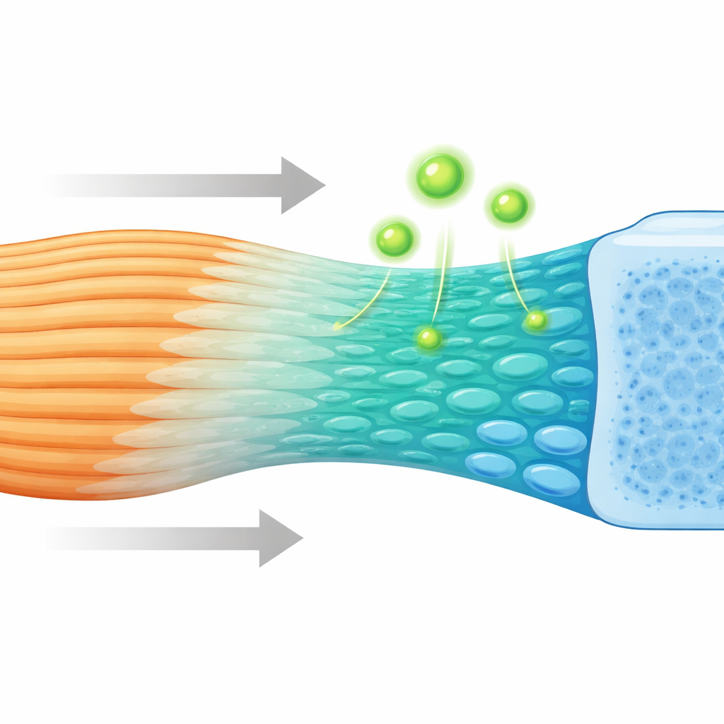
Where a tendon anchors into bone, the tissue does not change abruptly from soft to hard. Instead, it passes through a thin, graded layer of fibrocartilage that gradually shifts in composition and stiffness. This gradient helps smooth out stress and prevent tearing. While scientists have long suspected that specialized progenitor cells must build this zone, the identity and behavior of those cells have remained unclear. Using high-resolution spatial gene mapping and single-cell sequencing in mice, the researchers charted how thousands of individual cells are arranged and which genes they switch on as the attachment forms and matures.
Discovery of a Specialized Builder Cell
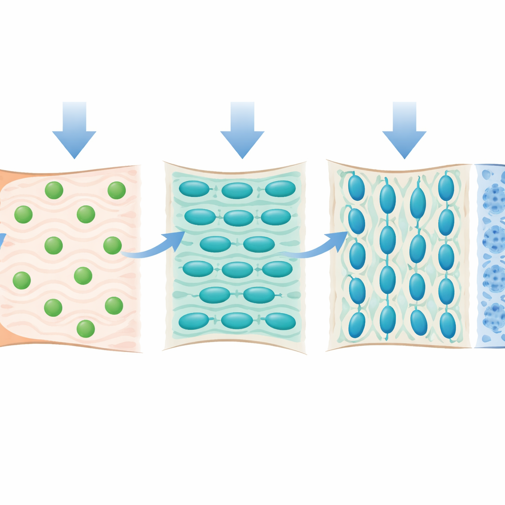
By following gene activity from late embryonic life through the first month after birth, the team identified a distinct population of progenitor cells marked by the gene Tnn (which encodes the matrix molecule tenascin-W). These Tnn-positive cells appear early at the tendon–bone interface, in a narrow band separate from ordinary tendon cells and from the cartilage of the nearby bone end. Lineage-tracing experiments showed that Tnn-marked cells and their descendants remain concentrated in the fibrocartilage region and are closely associated with zones that will later mineralize. Over time, these cells shift from a flexible, stem-like state toward a committed role in producing cartilage and mineral-rich matrix, essentially acting as dedicated builders of the tendon attachment fibrocartilage.
What Happens When These Builders Are Lost
To test whether Tnn-positive progenitors are truly necessary, the researchers created mice in which these cells could be selectively eliminated after birth. When Tnn-positive cells were ablated, the fibrocartilage at the tendon attachment developed abnormally. The normally layered structure became thin and disorganized, with fewer and smaller fibrocartilage cells. Imaging at microscopic and three-dimensional scales revealed reduced mineral content and weaker subchondral bone under the attachment zone. Mechanical testing confirmed that these altered entheses were less stiff and had a lower material strength, indicating that loss of the progenitor population leads to a structurally and functionally inferior tendon-to-bone connection.
How Load and Movement Shape the Interface
The study also asked how mechanical forces influence these progenitor cells. The investigators used botulinum toxin to partially paralyze a shoulder muscle, greatly reducing the normal pull on the tendon during growth. Under these unloaded conditions, the fibrocartilage remained underdeveloped: cells were smaller, the matrix was thinner, and key cartilage components such as type II collagen were markedly reduced. Single-cell analysis showed that the number of Tnn-positive progenitors dropped, and those that remained displayed a lower capacity to mature into cartilage-producing cells. Genes involved in matrix building, mineralization, and mechanosensing ion channels were also dampened, suggesting that the Tnn-positive cells are tuned to feel and respond to mechanical cues.
What This Means for Healing and Repair
In plain terms, this work shows that a transient, early wave of Tnn-positive progenitor cells specifically builds the fibrocartilage that anchors tendon to bone, and that normal mechanical loading is essential both to maintain their numbers and to unlock their cartilage-forming potential. When these cells are removed, or when muscle pull is taken away, the attachment zone stays stunted and mechanically weak. These insights help explain why tendon-to-bone repairs can fail and point toward future strategies: therapies may need to both recruit or protect specialized progenitors at the enthesis and provide appropriate mechanical stimulation to guide them, in order to truly regenerate a durable, graded tendon-to-bone connection.
Citation: Zhang, T., Zhang, L., Yuan, Z. et al. Identification of a new population of Tnn+ progenitors to form tendon enthesis fibrocartilage. Bone Res 14, 43 (2026). https://doi.org/10.1038/s41413-026-00519-3
Keywords: tendon enthesis, fibrocartilage, progenitor cells, mechanical loading, tissue regeneration