Clear Sky Science · en
Brain parcellation for TMD neuroimaging: a critical narrative review
Why jaw pain and brain maps matter
Many people live with long‑lasting pain in the jaw joint and the muscles used for chewing, a group of problems known as temporomandibular disorders (TMD). For some, scans of the jaw itself do not fully explain why the pain persists or why it interferes with mood, sleep, and daily life. This review article looks upward—from the jaw to the brain—to ask how modern brain‑imaging methods can better map the networks that shape TMD pain, and how choosing the right kind of brain map, or “parcellation,” can make those scans more reliable and more useful for future treatments.
Looking for answers inside the brain
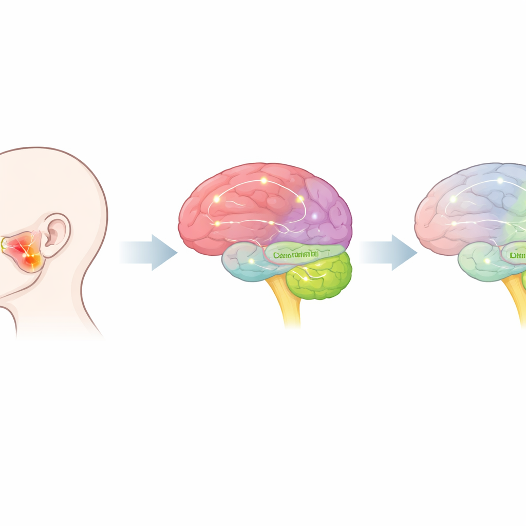
Over the past decade, brain scans have revealed that TMD is not just a problem of joints and muscles. Techniques such as functional MRI, which tracks changes in blood oxygen as a stand‑in for neural activity, and diffusion imaging, which traces major wiring pathways, repeatedly show changes in brain areas that feel, control, and regulate pain. These include regions that register touch and movement, areas that color pain with emotion, and deeper centers in the brainstem that can either dampen or amplify painful signals coming from the face. Because these changes are spread out across the brain, researchers need a clear way to divide the brain into named regions so they can compare results across studies.
Why carving up the brain is not straightforward
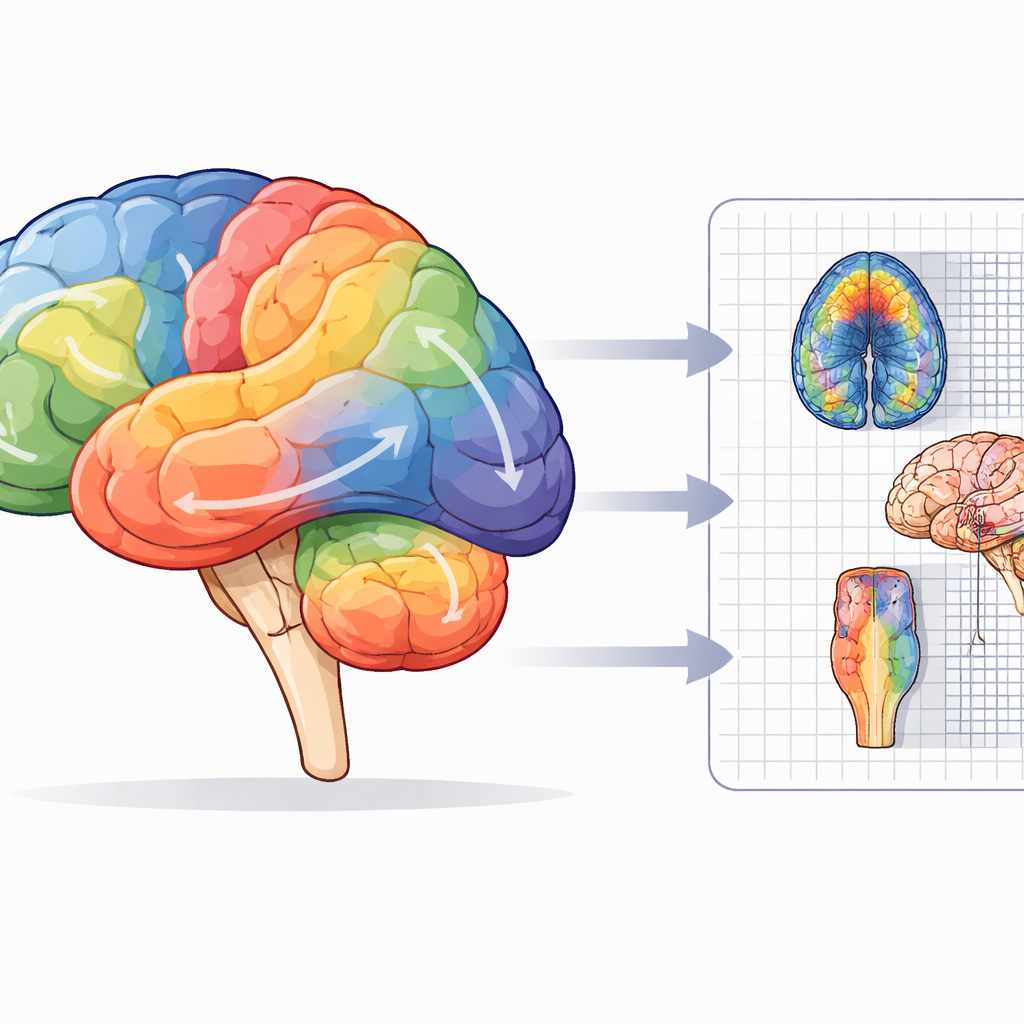
Brain parcellation is the process of slicing the brain into many small regions, a bit like turning a city map into neighborhoods and blocks. Some atlases follow visible anatomy, drawing borders along the folds and grooves of the brain surface. Others group together spots that are active at the same time, revealing functional networks such as those involved in movement, attention, or daydreaming. Still others blend several data sources at once. This review shows that no single atlas is ideal for every question. Atlases that are too coarse can hide important differences between nearby regions involved in face sensation and jaw movement. Atlases that are too fine can be noisy or hard to align across people and scanners. Researchers also need coverage that reaches beyond the outer brain surface into the cerebellum and brainstem, which play key roles in coordination and in turning pain volume up or down.
Choosing the right tools for jaw‑related pain
The authors compare widely used brain atlases and describe what each is good for in TMD research. Mid‑sized anatomical schemes, such as those that divide the cortex into major folds, are easy to interpret and work well for broad summaries, but they miss smaller hot‑spots that may be crucial for chronic pain. High‑detail atlases developed from large projects can pinpoint fine‑grained areas in the thinking and feeling parts of the brain, supporting precise network analyses. Other atlases are tailored to the cerebellum, improving the view of regions that help coordinate jaw movement and adapt to ongoing pain. Specialized brainstem maps zoom in on tiny nuclei that send powerful “stop” or “go” signals to incoming pain. The review also notes resources that help standardize large‑scale networks or validate how well automated software finds structures in the first place.
Putting the brain puzzle together
Because TMD affects many connected systems at once—sensation, movement, emotion, attention, and deep pain‑control loops—the authors argue that researchers should not rely on a single map. Instead, they propose a combined strategy: use a detailed multimodal atlas for the outer cortex, a specialized template for the cerebellum, and high‑resolution atlases for brainstem nuclei. Optional network‑focused or connectivity‑based atlases can be added when questions center on resting‑state networks or long‑range wiring. All of these should be brought into a common coordinate space and checked for consistency so that findings from different studies can be meaningfully compared.
What this means for patients and future care
In plain terms, the article concludes that better “cartography” of the brain will sharpen our view of how TMD alters pain circuits, from the jaw to the deepest brain centers. By carefully choosing and combining brain atlases, scientists can more reliably link certain patterns of activity or structure to symptoms, track changes over time, and test how treatments—from splints and exercises to medications or brain‑based therapies—reshape these networks. While this review does not test new treatments itself, it lays out a blueprint for more accurate and standardized brain imaging in TMD, a necessary step toward turning complex scans into clear guidance for diagnosis, prognosis, and personalized pain management.
Citation: Savychuk, N., Pekhno, V., Liakhovska, A. et al. Brain parcellation for TMD neuroimaging: a critical narrative review. BDJ Open 12, 39 (2026). https://doi.org/10.1038/s41405-026-00407-2
Keywords: temporomandibular disorders, brain imaging, pain networks, brain atlases, functional MRI