Clear Sky Science · en
Loss of regional theta differentiation in TMS-EEG response marks network dysfunction in psychosis risk
Why brain rhythms matter for mental health
Psychosis often seems to arrive suddenly, but the brain usually sends out early warning signs long before a first episode. This study asks whether tiny, millisecond-fast brain rhythms can reveal when communication between brain regions starts to go awry in young people at clinical high risk for psychosis. By gently nudging the brain with magnetic pulses and recording its electrical response, the researchers look for patterns that distinguish healthy brain networks from those that may be struggling but are not yet in full-blown illness.
Poking the brain to watch it react
The team worked with 44 young people seeking help who were classified as being at clinical high risk for psychosis and 58 healthy volunteers. All participants wore an electrode cap to record brain activity while receiving brief magnetic pulses to three areas on the surface of the brain: two regions in the frontal lobe involved in planning and self-related thinking, and one in the parietal lobe involved in attention and linking senses to actions. This combined method, called TMS-EEG, lets scientists trigger a small burst of activity in a chosen spot and then watch how the resulting waves spread across the brain over a fraction of a second.
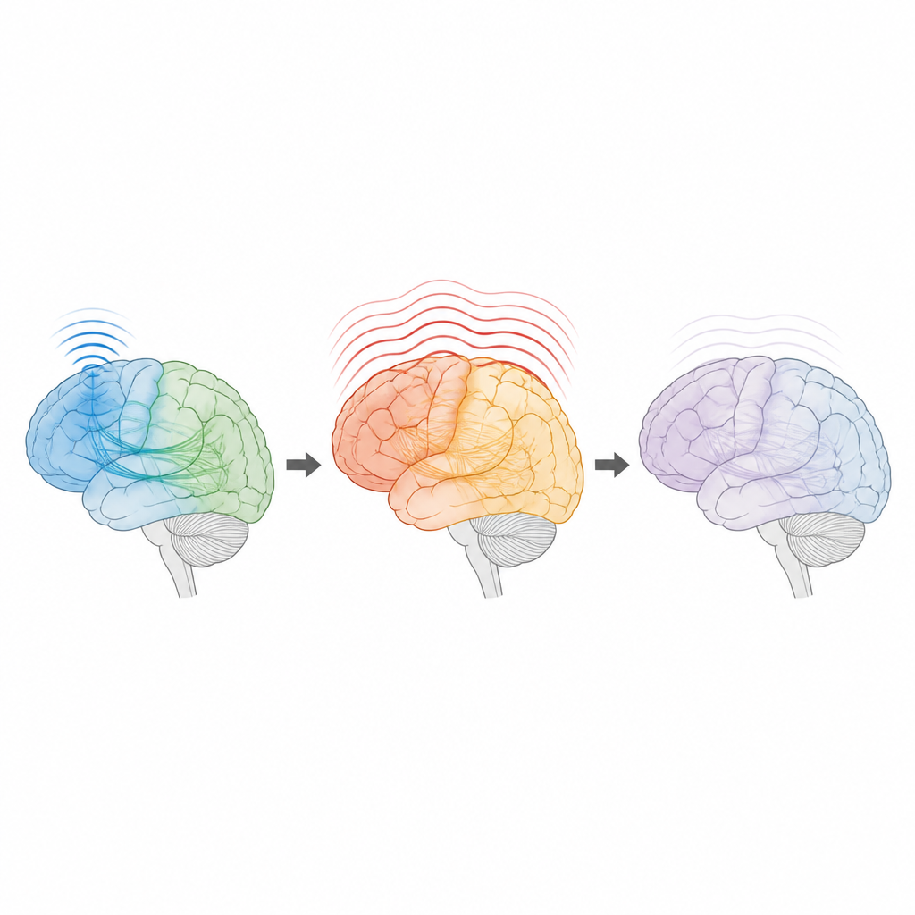
Slow brain waves that link distant regions
When neurons fire together they produce rhythmic signals, often referred to as brain waves. In this study the authors focused on two ranges: relatively slow theta waves and faster gamma waves. Theta waves are thought to coordinate long-distance communication between brain regions, while gamma waves seem more tied to local processing. After each magnetic pulse, the researchers measured how strongly these rhythms emerged over front and central parts of the head, and whether the pattern depended on which brain area was stimulated.
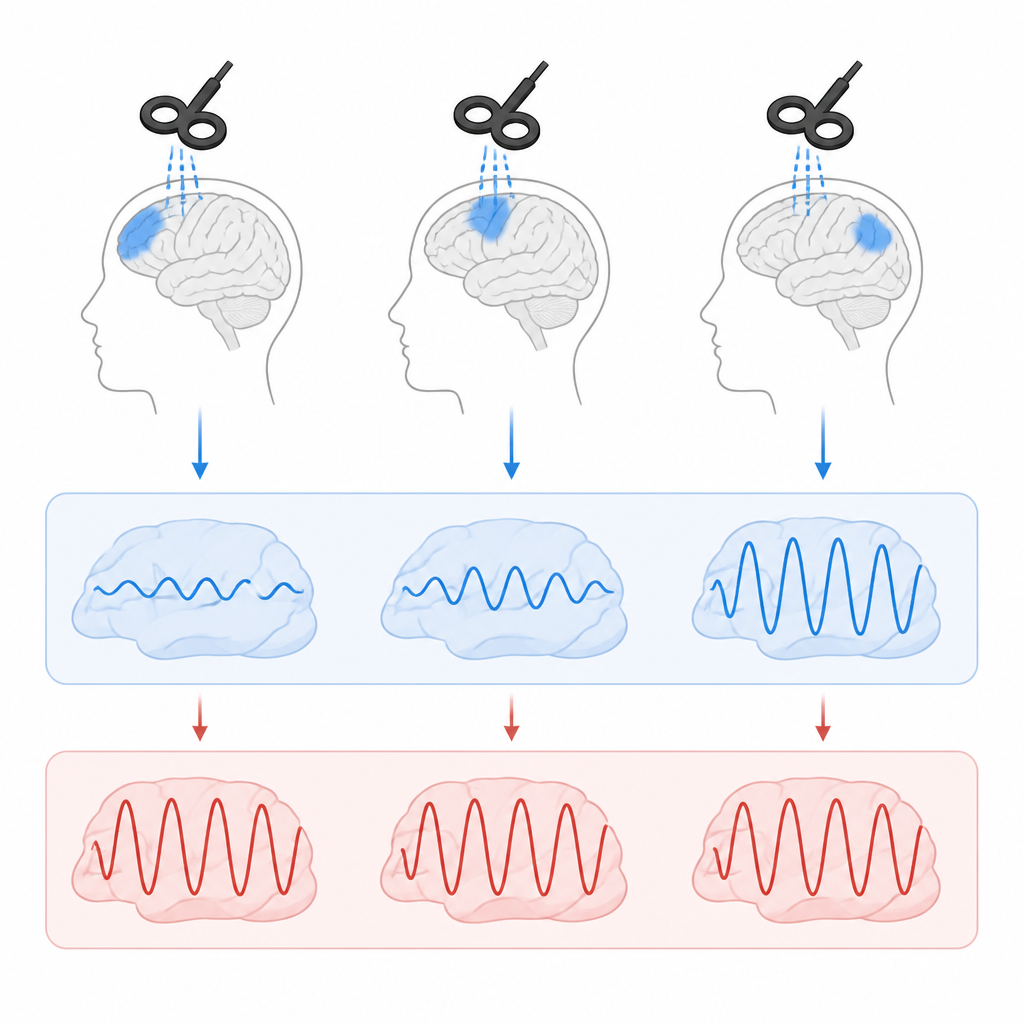
Healthy brains show a clear map, at-risk brains blur it
In healthy volunteers, the brain’s response in the theta range depended strongly on where the pulse landed. Stimulating one frontal area produced the strongest theta burst, while the parietal and midline frontal targets produced weaker or differently timed responses. This “fingerprint” suggested that each region and its connected network had its own characteristic way of reacting. In contrast, people at clinical high risk did not show this regional signature. No matter which of the three sites was stimulated, their theta responses looked similarly strong and lacked the clear differences seen in healthy brains.
Compensation instead of simple failure
The absence of regional differences might sound like a straightforward loss of function, but the story is more nuanced. The at-risk group did not simply show weaker signals. Instead, their theta responses were often larger and more uniform across sites, especially compared with healthy volunteers. Importantly, within the high-risk group, stronger theta responses in certain regions were linked to fewer unusual thoughts, more emotional experience, and better everyday role functioning. This pattern hints that the brain may be turning up these long-range rhythms to compensate for underlying structural problems in its wiring, at least for a time.
What the study did not find
Earlier research in people with established psychotic disorders often found disrupted gamma activity after brain stimulation. In this sample of high-risk individuals, however, gamma responses did not differ reliably from healthy controls, and their ties to symptoms were weak and inconsistent. This suggests that changes in the slower theta rhythms may appear earlier in the course of illness, while more dramatic alterations in faster rhythms could emerge closer to or after a first psychotic episode.
How this helps us understand psychosis risk
To a layperson, the key message is that the brains of people at high risk for psychosis may still be working hard to hold things together. Instead of distinct regions each doing a specialized job, their networks appear to respond in a more uniform way, as if recruiting extra help from neighboring systems. Because higher theta responses are linked to milder symptoms, this “all hands on deck” pattern may reflect a temporary safety net before the system reaches a tipping point. While this single study cannot predict who will become ill, it shows that noninvasive brain stimulation and recording can reveal subtle changes in communication between brain regions, offering a promising path toward earlier and more precise assessments of psychosis risk.
Citation: Zimmermann, N., Liebrand, M., Michel, C. et al. Loss of regional theta differentiation in TMS-EEG response marks network dysfunction in psychosis risk. Transl Psychiatry 16, 255 (2026). https://doi.org/10.1038/s41398-026-04030-5
Keywords: psychosis risk, brain oscillations, TMS-EEG, theta rhythms, brain networks