Clear Sky Science · en
Amyloid fibril polymorphism in the heart and liver of a patient with polyneuropathic ATTRv-V122Δ amyloidosis
Why strange protein clumps matter
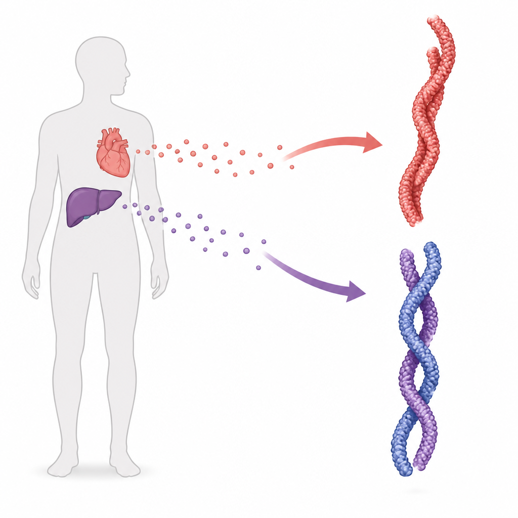
Many serious diseases arise when normal proteins in our bodies twist into stubborn fibers that the body cannot clear. In this study, researchers examined such fibers from the heart and liver of one man with a rare inherited form of transthyretin amyloidosis, a condition that can damage nerves and the heart. By zooming in with cryo electron microscopy, they discovered that these fibers come in more than one shape, hinting that the physical form of these protein clumps may help explain why symptoms vary so much between patients.
A protein that travels and sometimes misbehaves
Transthyretin is a transport protein made mainly in the liver that normally helps carry thyroid hormone and vitamin A in the blood and other body fluids. In transthyretin amyloidosis, the protein loses its usual form and stacks into long, rigid fibers called amyloid. These fibers can build up in many organs, including the heart, nerves, eyes, and gut. Some people develop mainly heart problems, others mostly nerve damage, and some show a mix of both, and over 220 genetic variants of transthyretin have been linked to these diverse patterns.
A rare deletion and a close look inside tissues
The patient studied carried a rare deletion of a single building block in the transthyretin gene, known as V122Δ, and mainly suffered from nerve-related problems along with heart involvement. After his death, the team obtained samples from his heart and liver. Stains that highlight amyloid confirmed heavy deposits in the heart muscle, nearby nerves, blood vessel walls, and surrounding fat. The scientists then gently extracted the fibers and used several laboratory methods to verify that they were made of transthyretin fragments typical of one common fibril type found in this disease.
One protein, multiple fiber shapes
Using cryo electron microscopy, which can reveal structures near the atomic scale, the researchers imaged thousands of individual fibers. In both heart and liver, they saw a mix of straight and twisted fibers, but most were twisted. Detailed reconstruction showed that many fibers consisted of a single strand, while a substantial fraction formed double strands, where two protofilaments wound around each other. In all of these forms, each strand had a core fold closely matching that seen in other transthyretin amyloidosis patients, including those with heart-dominant disease and those with other mutations.
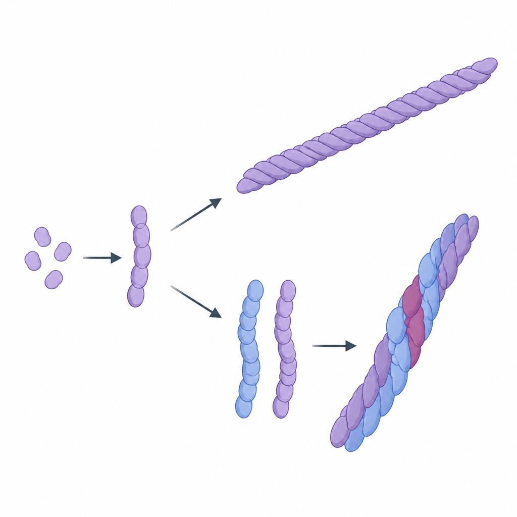
Subtle differences at contact points
Although the basic fold was conserved, the way strands associated in the double fibers differed from forms described in other organs and mutations. In one double form, two strands contacted each other in an uneven way, with specific amino acids forming bridges between them. In another, the pair showed a more symmetric relation, again held together by defined contacts. Intriguingly, regions that form a narrow internal channel within the fiber, previously suspected to differ between disease types, looked similar to the closed state seen in mainly heart-based disease, suggesting that other aspects of the double-strand arrangement might matter for symptoms.
Possible links to symptoms and treatment
These observations add to a growing picture in which patients with mostly heart problems tend to show a single, uniform fiber structure in cardiac tissue, while those with strong nerve involvement, like carriers of the V122Δ and I84S variants, show a richer mix of fiber forms. The authors propose that this structural variety could help explain why some patients develop nerve damage, although current data from only one V122Δ patient cannot settle the issue. Present drugs for transthyretin amyloidosis mainly work by stabilizing the normal protein or reducing its production, so they likely act before fibers form. However, if future therapies aim to clear amyloid deposits directly, they will need to account for these different shapes or focus on regions that are shared across them.
What this work means for patients
In simple terms, this study shows that in one person with nerve-heavy transthyretin amyloidosis, the harmful protein fibers in the heart and liver were not all alike: some were single strands, others were double, and they could even switch shapes along the same fiber. While more patients must be studied, this structural variety may be one piece of the puzzle behind the wide range of symptoms seen in this disease. Mapping these shapes more completely could guide better diagnostic tools and help future treatments home in on common weak points in the fibers, regardless of which organs they affect.
Citation: Ahmed, Y., Nguyen, B.A., Kelly, C. et al. Amyloid fibril polymorphism in the heart and liver of a patient with polyneuropathic ATTRv-V122Δ amyloidosis. Commun Biol 9, 713 (2026). https://doi.org/10.1038/s42003-026-09919-x
Keywords: transthyretin amyloidosis, amyloid fibrils, polyneuropathy, heart and liver, cryo electron microscopy