Clear Sky Science · en
Glucocorticoids induce a phagocytic C1Q+ macrophage phenotype primed for IFNγ-dependent CXCL9 secretion
Why stress hormones in cancer matter
Many people know glucocorticoids as stress or anti-inflammatory hormones given as medications like cortisone. This study looks at how these hormones behave inside a rare adrenal cancer called adrenocortical carcinoma and how they change the behavior of immune cells that live in and around tumors. The findings challenge the simple idea that these hormones always weaken anti-cumor immunity and instead reveal a more nuanced picture that could help doctors better use immunotherapy.
The setting inside adrenal tumors
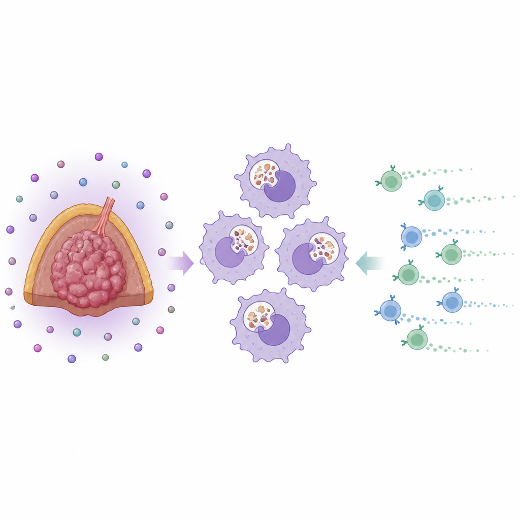
Adrenocortical carcinoma arises in the adrenal glands, which naturally produce large amounts of steroid hormones. Because many of these tumors secrete extra cortisol, they are a natural testbed for understanding how stress hormones shape the local immune landscape. The researchers examined human tumor samples and found that these cancers are heavily populated by a type of white blood cell called macrophages. Most of these cells carried markers known as CD68 and CD163, a combination usually linked to a healing or tissue-remodeling profile rather than an aggressively inflammatory one. Importantly, the number and basic type of these macrophages did not simply track with how much cortisol the tumor produced, the tumor stage, or the patient’s sex.
Hormone-shaped clean-up cells
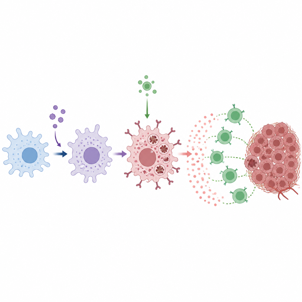
To understand what glucocorticoids do to macrophages in more detail, the team recreated the process in the lab. They took blood cells from healthy donors and exposed developing macrophages to either standard immune signals or the same signals plus a synthetic glucocorticoid. With hormone exposure, the cells strongly turned on genes and proteins for CD163 and a molecule called C1q. C1q helps immune cells recognize and engulf dying cells, acting like a molecular handle on their surface. When macrophages were grown in liquid taken from hormone-producing cancer cell lines, they adopted the same C1q-rich, CD163-rich profile, and blocking steroid production in the cancer cells largely prevented this shift. Gene analyses and functional tests then showed that these hormone-conditioned macrophages were especially good at swallowing dying cancer cells, relying heavily on C1q to do so.
From clean-up to T cell recruitment
Macrophages do more than clear debris; they also call in other immune cells. The researchers found that when these C1q-high macrophages were later exposed to interferon gamma, a signal commonly boosted during immune checkpoint therapy, they released large amounts of a T cell-attracting chemical called CXCL9. In fact, they secreted more CXCL9 than classical pro-inflammatory macrophages. This effect depended on functional glucocorticoid receptors, because blocking these receptors with the drug mifepristone reversed the special macrophage state and sharply reduced CXCL9 release. Across tumor datasets, higher expression of macrophage and C1q genes went hand in hand with stronger markers of T cell presence and better patient survival, suggesting that this CXCL9-producing macrophage subset may favor a more effective anti-tumor response.
Animal and patient clues about treatment
The team then explored how these insights play out during immunotherapy. In mice bearing glucocorticoid-producing adrenal tumors, immune checkpoint treatment slowed tumor growth and increased both CXCL9 levels in the tumor and the presence of CD4 and CD8 T cells. However, when the same mice also received mifepristone, the benefit of checkpoint therapy was blunted, CXCL9 staining in tumors dropped, and CD4 T cell numbers fell. In patients with adrenal cancer receiving checkpoint inhibitors, blood levels of CXCL9 rose after treatment, while T cells carrying the matching CXCR3 receptor tended to disappear from the bloodstream, consistent with their movement into tissues. Patients whose tumors contained more CD163-positive macrophages at surgery were more likely to later respond to immunotherapy than those with fewer such cells.
What this means for future care
For non-specialists, the key message is that stress hormones inside tumors do not simply shut immunity down. In adrenal cancer, they can reprogram local macrophages into highly active clean-up cells that, once triggered by immune signals, send out strong beacons to attract T cells. These hormone-shaped macrophages may help checkpoint drugs work better, and their abundance in tumor samples could serve as a useful sign of which patients are more likely to benefit. At the same time, broadly blocking glucocorticoid signals in such tumors might unintentionally weaken this helpful arm of the immune response, suggesting that future treatments must carefully balance hormone control with support for beneficial immune cells.
Citation: Triebig, A.S., Maier, T., Schwarzlmueller, P. et al. Glucocorticoids induce a phagocytic C1Q+ macrophage phenotype primed for IFNγ-dependent CXCL9 secretion. Sci Rep 16, 15345 (2026). https://doi.org/10.1038/s41598-026-52733-y
Keywords: adrenocortical carcinoma, glucocorticoids, tumor macrophages, CXCL9, immune checkpoint therapy