Clear Sky Science · en
Dysregulated B cell homeostasis and the involvement of B cell-activating factor in the progression of diabetic retinopathy
Why this study matters for people with diabetes
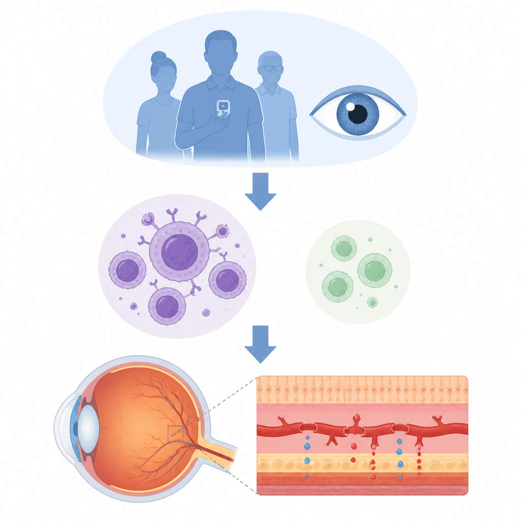
People with diabetes often worry about losing their sight, yet the invisible battles inside the eye that lead to vision loss remain hard to picture. This study looks at how a specific branch of the immune system, called B cells, and a signaling molecule that nurtures them, may quietly weaken the eye’s delicate blood vessels and promote diabetic retinopathy, a leading cause of blindness in adults.
The eye disease behind blurry and lost vision
Diabetic retinopathy develops when long term high blood sugar damages the tiny blood vessels in the retina, the light sensing tissue at the back of the eye. Early on, vessels become leaky and the blood–retina barrier, which normally keeps the eye’s environment tightly controlled, starts to fail. In advanced stages, abnormal new vessels grow and scar tissue forms, threatening severe and permanent vision loss. Inflammation has long been known as a key driver of this process, but most attention has focused on other immune cells, leaving the role of B cells relatively unexplored.
Immune cells out of balance in patients and mice
The researchers examined blood and eye fluid from people with different stages of diabetic retinopathy and compared them with healthy individuals. They found that patients with the proliferative, most advanced form of the disease had more B cells overall but far fewer of a specialized calming subset known as regulatory B cells. At the same time, levels of a protein called BAFF, which supports B cell growth and survival, were significantly higher in both blood and the clear fluid at the front of the eye. These shifts point to an immune system tilted toward activation rather than control, especially in advanced disease.
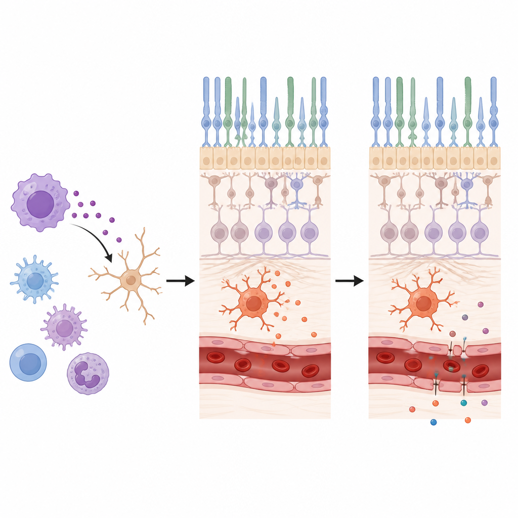
High sugar and BAFF stir up brain like cells in the retina
To see whether these immune changes also occur in a controlled setting, the team turned to a well established mouse model of diabetes. Diabetic mice showed the same pattern as patients: an expansion of total B cells, a drop in regulatory B cells, and a strong rise in BAFF within the retina itself. In parallel cell culture experiments, exposing retinal microglia, the eye’s resident brain like immune cells, to high sugar levels boosted their production of BAFF and its main receptor. When the scientists added extra BAFF, the microglia became more activated, a state associated with releasing inflammatory factors that can harm nearby blood vessels and nerve cells.
Blocking the signal reduces leaky retinal vessels
The group then asked whether dampening BAFF could protect the retina from diabetes related damage. Diabetic mice were treated over several months with a BAFF neutralizing antibody. Standard eye imaging did not show dramatic differences in vessel structure between treated and untreated diabetic animals, which is expected in this relatively early disease model. However, a sensitive dye based test revealed that diabetic mice had much leakier retinal vessels than healthy controls, and that blocking BAFF significantly reduced this leakage. This suggests that BAFF is closely linked to early blood–retina barrier breakdown, even before severe structural changes appear.
What these findings could mean for future care
Taken together, the study paints a picture of diabetic retinopathy as not only a disease of sugar damaged vessels, but also of misdirected B cells and an overabundant growth signal, BAFF, that fuels inflammatory activity in the retina. By shifting the balance away from protective regulatory B cells and toward more aggressive immune responses, this pathway appears to weaken the eye’s vascular shield and promote fluid leakage. While more work is needed, particularly in humans, targeting BAFF or restoring regulatory B cell function could one day complement current treatments by addressing the immune side of diabetic eye disease.
Citation: Wang, Y., Cui, L., He, W. et al. Dysregulated B cell homeostasis and the involvement of B cell-activating factor in the progression of diabetic retinopathy. Sci Rep 16, 15451 (2026). https://doi.org/10.1038/s41598-026-46863-6
Keywords: diabetic retinopathy, B cells, BAFF, retinal inflammation, blood retina barrier