Clear Sky Science · en
Multiplexed optical barcoding and sequencing for spatial omics
Seeing Where Molecules Live in Tissues
Our bodies are made of many kinds of cells packed together in precise patterns, and those patterns matter for health and disease. Scientists have powerful tools to measure which genes are active in cells, but these tools often lose track of where in the tissue each measurement came from. This paper introduces a new method, called MOLseq, that aims to keep both pieces of information at once: what each cell is doing and exactly where it is located.
Why Location Matters for Molecules
In recent years, “spatial omics” has changed how researchers look at tissues. Instead of studying cells in isolation, scientists now ask which genes are switched on in each cell while that cell remains in its original neighborhood. Existing approaches fall into two main camps. Imaging methods use microscopes and fluorescent tags to directly see thousands of molecules in cells with very fine detail, often down to subcellular structures. But they usually require scientists to pick which genes to look at in advance. Sequencing methods, in contrast, can read out virtually all active genes at once, but they typically smear together signals from many cells and only record positions in coarse two‑dimensional grids. As a result, researchers often must choose between breadth of information and sharpness of location.
A Light-Controlled Address System
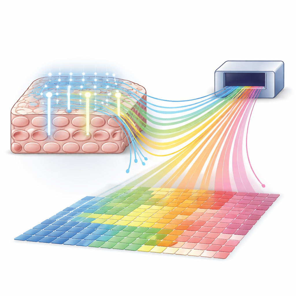
MOLseq offers a way to combine the breadth of sequencing with the fine control of light. The core idea is to give molecules inside cells a kind of “postal code” that records their position, then read both the postal code and the gene identity by sequencing. First, the method attaches a short DNA primer to messenger RNAs inside fixed cells and converts these RNAs into DNA copies. Then, using a projector-like device that shines patterned ultraviolet (UV) light, MOLseq adds short DNA “letters” to these copies only in illuminated regions. Each burst of light adds exactly one letter, and the sequence of letters builds a unique barcode for each location. After several rounds, molecules from different regions carry different barcodes, which act as their spatial addresses when the sample is later broken apart and sequenced.
Because barcodes are built step by step, the number of possible addresses grows rapidly with the number of letters and rounds. The authors show that their light-driven chemistry can add letters in series with about 90% success per step, and that multiple letters can be managed in parallel using designed helper strands. In cell culture experiments, they generated hundreds of distinct barcodes in situ and confirmed the expected barcode lengths using a DNA sizing instrument. Importantly, they also designed barcode schemes that can flag and correct some errors, improving reliability even when individual steps are not perfect.
Writing Barcodes One Cell at a Time
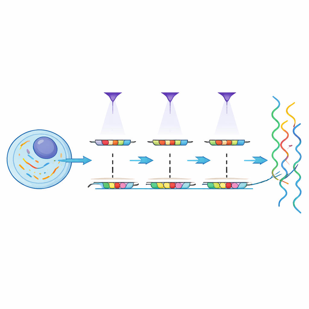
A key promise of MOLseq is precise control over where barcodes are written. By steering UV light with a digital micromirror device, the team selectively photocleaved and ligated letters in regions ranging from large patches down to individual cells. They used fluorescent probes to visualize where letters had been added, showing that neighboring cells just a few micrometers away received almost no unintended signal. In one experiment, they successfully assigned unique three-letter barcodes to 64 individual cells in the same dish. Modeling of the data indicated that the chance of an off-target letter being added in any given round was only a few percent, while the intended on-target addition rate remained high.
To test whether these barcodes can guide full gene readouts, the researchers mixed human and mouse cells in separate regions on the same coverslip and applied MOLseq. They built distinct two-letter barcodes for the human and mouse regions, confirmed their spatial separation with imaging, and then sequenced the barcoded material. Reads carrying the “human-region” barcode overwhelmingly mapped to human genes, and those with the “mouse-region” barcode to mouse genes. The small fraction of apparent mix-ups was similar to what is expected from natural sequence similarity between the two species, suggesting that most errors did not arise from the barcoding itself but from unavoidable ambiguities in read mapping.
Promise and Next Steps
By merging light patterning with DNA sequencing, MOLseq points toward a future where scientists can scan large tissue areas, capture the activity of many genes without preselection, and still know where each signal came from—potentially down to single cells. The current version still faces challenges: off-target light effects, limited efficiency with many ligation rounds, and the difficulty of capturing enough RNA from very small regions. Yet the study shows that multiplexed optical barcoding is practical and accurate in cultured cells, and it outlines realistic paths to scale up barcode diversity and error correction. For readers, the takeaway is that tools like MOLseq may soon let researchers create detailed “molecular maps” of tissues, revealing how cells’ positions and gene activity work together in development, brain function, cancer, and many other biological processes.
Citation: Venkatramani, A., Ciftci, D., Pham, K. et al. Multiplexed optical barcoding and sequencing for spatial omics. Sci Rep 16, 14086 (2026). https://doi.org/10.1038/s41598-026-41186-y
Keywords: spatial omics, optical barcoding, transcriptomics, single-cell profiling, DNA sequencing