Clear Sky Science · en
Functional effects of EpCAM N-glycosylation in MDA-MB-468 breast cancer cells
Why tiny sugar tags on cancer cells matter
Most of us think of sugar as something in our diet, but cells also use complex sugar chains as little decorations on their proteins. These sugar tags can change how proteins behave, and they are often altered in cancer. This study asks a focused question: do specific sugar tags on a well-known cancer marker called EpCAM actually change how aggressive breast cancer cells are, or do they mostly tweak the protein itself without reshaping the disease?
A marker on the cancer cell surface
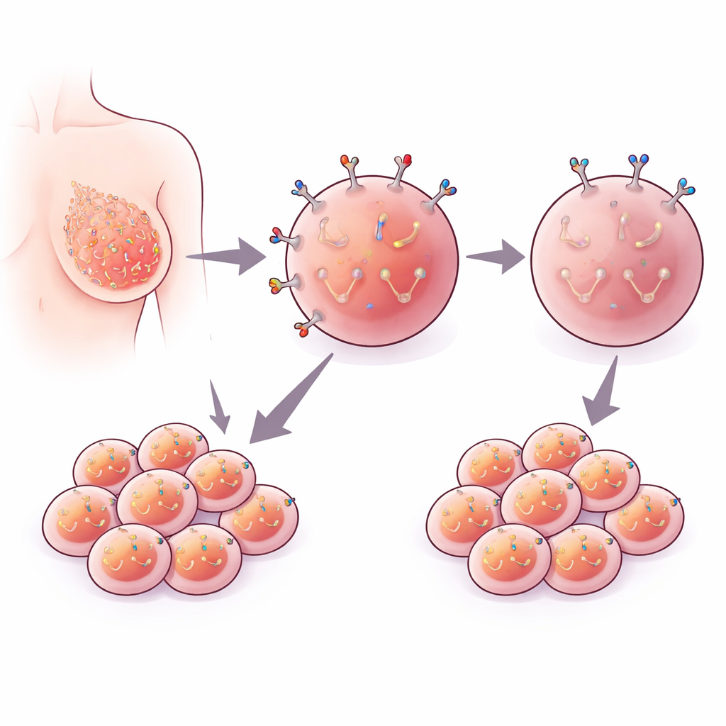
EpCAM is a protein that sits in the outer membrane of many epithelial cells, including breast cells. In several cancers, tumor cells carry much more EpCAM on their surface than healthy cells do, and patients whose tumors have high EpCAM levels often fare worse. Because of this, EpCAM has become a popular target for diagnostic tests and experimental therapies. Yet scientists still do not fully agree on whether EpCAM is actively driving cancer spread or merely tagging along as a bystander associated with other harmful changes.
Zooming in on EpCAM’s sugar coat
EpCAM naturally carries three small sugar attachments at precise positions in its structure. Earlier patient studies suggested that tumors with more EpCAM also showed more of certain sugar types, hinting that this sugar coating might influence how EpCAM behaves in metastasis. Directly turning off sugar addition on just one protein is technically difficult, so the researchers used gene engineering to sidestep this problem. Working in a triple-negative breast cancer cell line called MDA-MB-468, they first created cells in which the normal EpCAM gene was completely removed. They then put back either a normal, sugar-ready version of EpCAM or a subtly altered version in which the three sugar-accepting sites were changed so that sugars could no longer be attached.
What happens when the sugar tags are removed
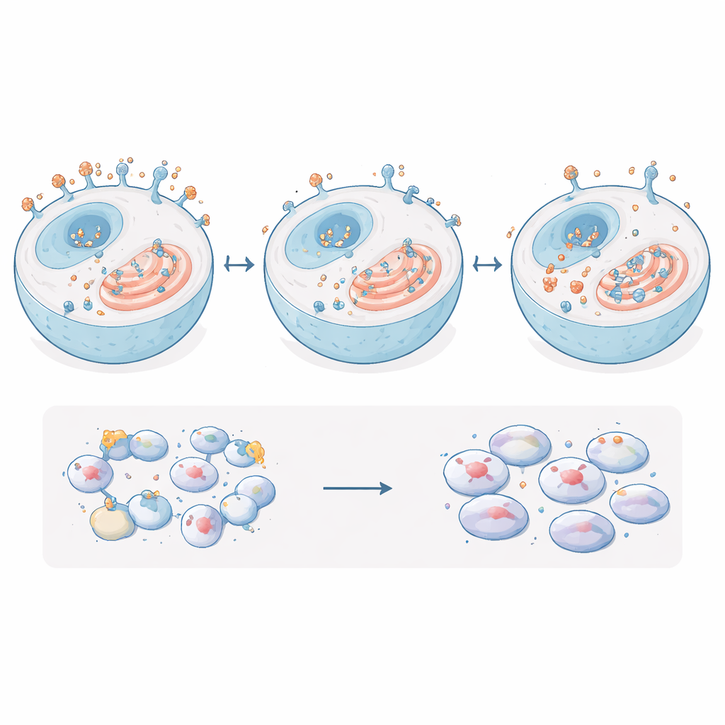
With this clean system in hand, the team measured what happened to the EpCAM protein itself. Removing the sugar sites made EpCAM much less stable: when cells were treated to stop new protein production, the sugar-free EpCAM degraded noticeably faster than normal EpCAM. Microscopy further showed that the sugar-free version did not reach the cell surface as efficiently. Instead, it tended to linger inside the cell, particularly in the endoplasmic reticulum, a folded internal membrane system where proteins are folded and checked for quality. Normal EpCAM, in contrast, was concentrated at the cell membrane where it can contact neighboring cells and interact with signaling partners.
What does not change inside the cancer cells
Because EpCAM has been linked to multiple growth and survival pathways, the researchers next asked whether stripping its sugar tags altered key downstream molecules or cancer-like behaviors. They measured a set of proteins and genes that are often controlled by EpCAM, including major growth regulators and adhesion molecules, and found no meaningful changes when comparing normal, EpCAM-lacking, EpCAM-overproducing, and sugar-free EpCAM cells. The team also carried out standard lab tests for traits tied to metastasis: how fast the cells grow, how well they move through a porous barrier, how readily they invade a gel that mimics surrounding tissue, and how strongly they stick to one another. All of these properties were essentially unchanged by the loss of EpCAM sugars. Only when EpCAM itself was heavily overproduced did they see a modest increase in how tightly the cells clumped together.
What this means for understanding and targeting EpCAM
Put simply, the study shows that the sugar coat on EpCAM strongly influences the protein’s stability and where it sits inside breast cancer cells, but it does not, by itself, switch these cells into a more or less aggressive state in this model. In MDA-MB-468 cells, removing EpCAM’s sugars makes the protein less durable and keeps more of it trapped inside the cell, yet basic behaviors linked to tumor spread remain largely the same. This suggests that while EpCAM and its sugar decorations are important pieces of the molecular puzzle, their functional impact may depend heavily on cell type and context. For therapy design, the work underscores that changing or targeting EpCAM’s sugar pattern may alter the protein itself without necessarily producing a dramatic, predictable change in how certain breast cancer cells behave.
Citation: Jenkinson, N.M., Oza, H., Yarema, K.J. et al. Functional effects of EpCAM N-glycosylation in MDA-MB-468 breast cancer cells. Sci Rep 16, 10021 (2026). https://doi.org/10.1038/s41598-026-38920-x
Keywords: EpCAM, breast cancer, cell adhesion, protein glycosylation, metastasis