Clear Sky Science · en
CD146 + interstitial cells contribute to the dystrophic skeletal muscle phenotype in vitro
Why muscle repair sometimes goes wrong
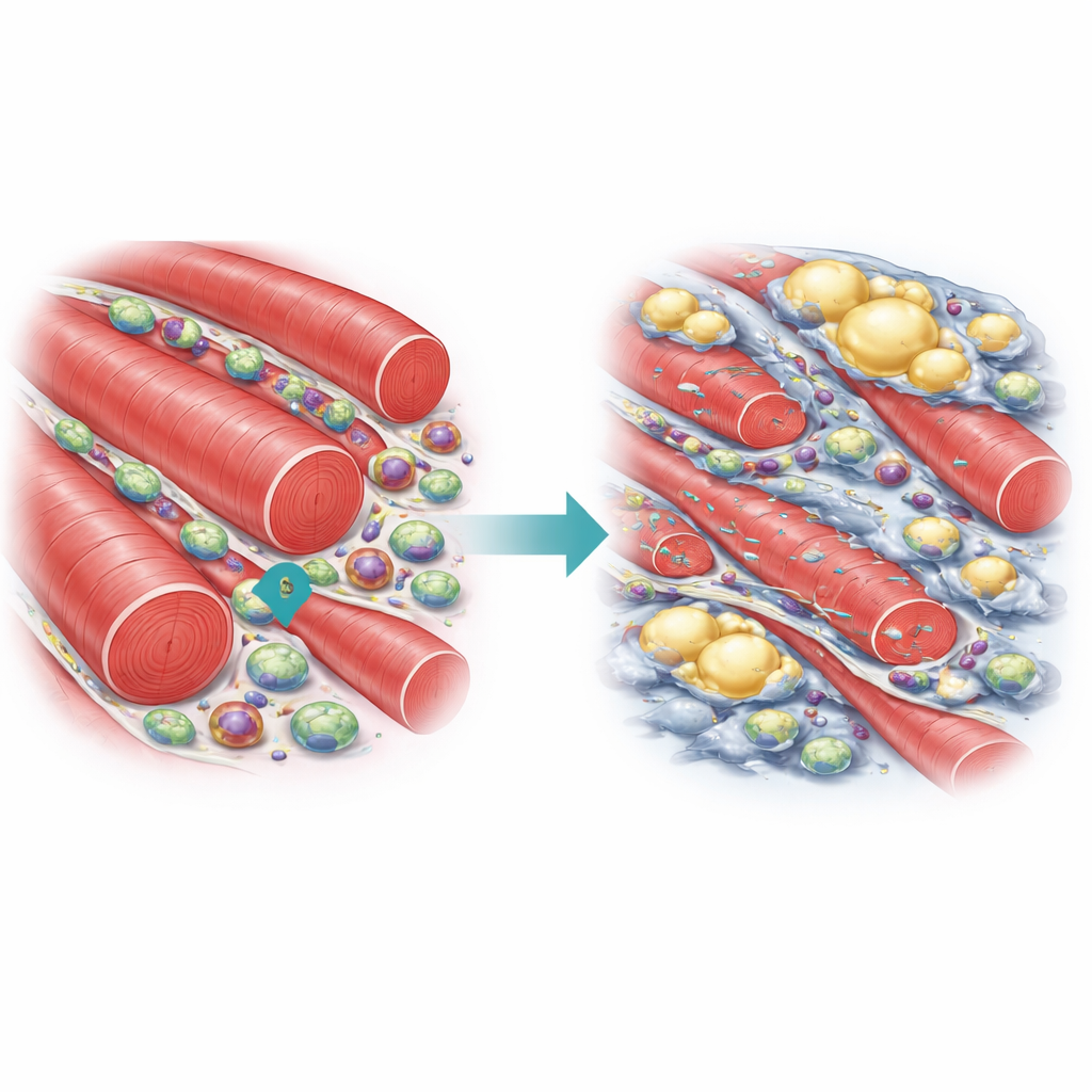
Duchenne muscular dystrophy is a severe childhood disease in which muscles gradually weaken and are replaced by scar and fat. Most research has focused on the muscle stem cells that normally rebuild damaged fibers. This study looks instead at a lesser-known group of support cells that sit around tiny blood vessels in muscle. By asking how these CD146-positive interstitial cells behave in healthy and diseased muscle, the authors uncover how they may quietly push dystrophic muscle toward scarring and poor blood supply instead of repair.
Hidden helpers in healthy muscle
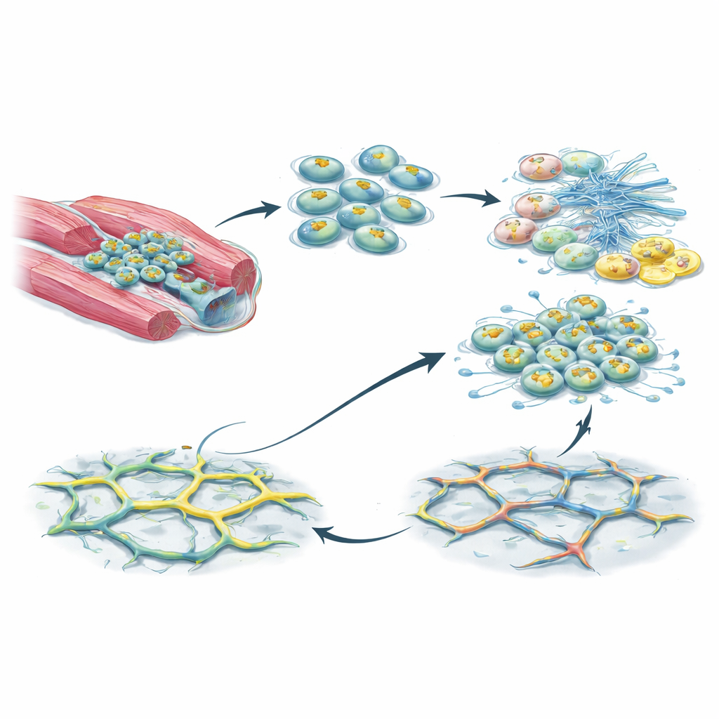
In normal muscle, many cell types cooperate to restore tissue after injury. Among them are pericyte-like cells that wrap around capillaries and can give rise to new muscle, connective tissue, or fat. These cells often carry a surface marker called CD146. In mice without disease, CD146-positive cells sit mostly along blood vessels, and past work has shown they can support regeneration by forming new muscle fibers and helping remodel the surrounding tissue. The authors started by mapping where these cells sit in mouse leg muscles and measuring how many of them also carry markers linked to blood vessel support or fibrous tissue production.
A different identity in dystrophic muscle
Using the mdx mouse, a standard model of Duchenne muscular dystrophy, the team found that muscles lacking dystrophin contained fewer CD146-positive pericyte-like cells overall. Those that remained were often located within fibrotic, scar-like regions rather than just around vessels. When the researchers isolated CD146-positive cells from healthy and mdx muscles and grew them in dishes, clear differences emerged. Cells from dystrophic muscle divided faster but were much less likely to form muscle fibers. Instead, they more readily became fibroblasts that lay down collagen, as well as fat cells filled with lipid droplets. Gene-expression profiling supported this shift: mdx CD146-positive cells turned down muscle-related genes and turned up genes tied to extracellular matrix, fibrosis, and tissue remodeling, resembling fibro–adipogenic progenitor cells more than classic muscle-forming progenitors.
How these cells may hinder blood vessel growth
Because muscle repair also depends on restoring a healthy blood supply, the authors tested whether the substances released by CD146-positive cells influenced vessel formation. They grew human endothelial cells on a gel that allows them to form capillary-like tubes and bathed them in medium that had previously been conditioned by mouse CD146-positive cells. Medium from healthy cells allowed fairly normal tube networks. In contrast, medium from mdx CD146-positive cells markedly reduced the number and length of tubes, indicating impaired angiogenesis. Measurements of secreted factors helped explain why: dystrophic cells produced less SDF-1 and angiopoietin-1, both known to attract and stabilize blood vessels, while releasing more angiopoietin-2, which can destabilize vessels when other growth signals are low.
Signals that push cells toward scarring
To probe the internal control systems of these cells, the researchers examined major signaling molecules that act as switches for inflammation, growth, and differentiation. Their RNA sequencing and protein analyses pointed to changes in NF-κB and AP-1 family members (c-Jun and c-Fos), networks known to be overactive in Duchenne muscles. In mdx CD146-positive cells, the activated form of NF-κB and c-Jun was elevated, while active c-Fos was reduced. These shifts match a pattern in which pro-inflammatory and pro-fibrotic programs are turned on, and muscle-building programs governed by factors like MyoD and myogenin are dialed down. Together, the data suggest that the dystrophic environment rewires these perivascular cells so they favor scar and fat formation and release signals that undermine blood vessel growth.
What this means for treating muscle disease
For non-specialists, the take-home message is that not all cells near a damaged muscle fiber are trying to help. In healthy muscle, CD146-positive pericyte-like cells can contribute to rebuilding fibers and supporting tiny vessels. In Duchenne muscular dystrophy, however, the same class of cells becomes more like scar-building and fat-forming cells and secretes factors that make it harder to form stable capillaries. By identifying the signaling pathways that drive this harmful shift, the work points to new treatment ideas: instead of targeting only muscle stem cells, future therapies might also aim to "re-educate" or restrain these interstitial cells, reducing fibrosis and improving blood flow so that any regenerative therapy has a better chance to succeed.
Citation: Mierzejewski, B., Michalska, Z., Kulma, D. et al. CD146 + interstitial cells contribute to the dystrophic skeletal muscle phenotype in vitro. Sci Rep 16, 10331 (2026). https://doi.org/10.1038/s41598-026-38311-2
Keywords: Duchenne muscular dystrophy, skeletal muscle regeneration, pericytes, fibrosis, angiogenesis