Clear Sky Science · en
Isotonic and minimally invasive optical clearing media for live cell imaging ex vivo and in vivo
Seeing Deeper into Living Tissues
Modern biology relies heavily on fluorescent microscopes to watch living cells and organs in action. Yet many of the most interesting events happen deep inside cloudy, opaque tissue, where light is quickly scattered and blurred. This paper describes a new way to gently make living mammalian tissues optically clearer, called SeeDB-Live, allowing scientists to see farther into brains, organoids and cell clusters without poisoning or disturbing the cells they want to study.
Why Living Tissues Are Hard to See Through
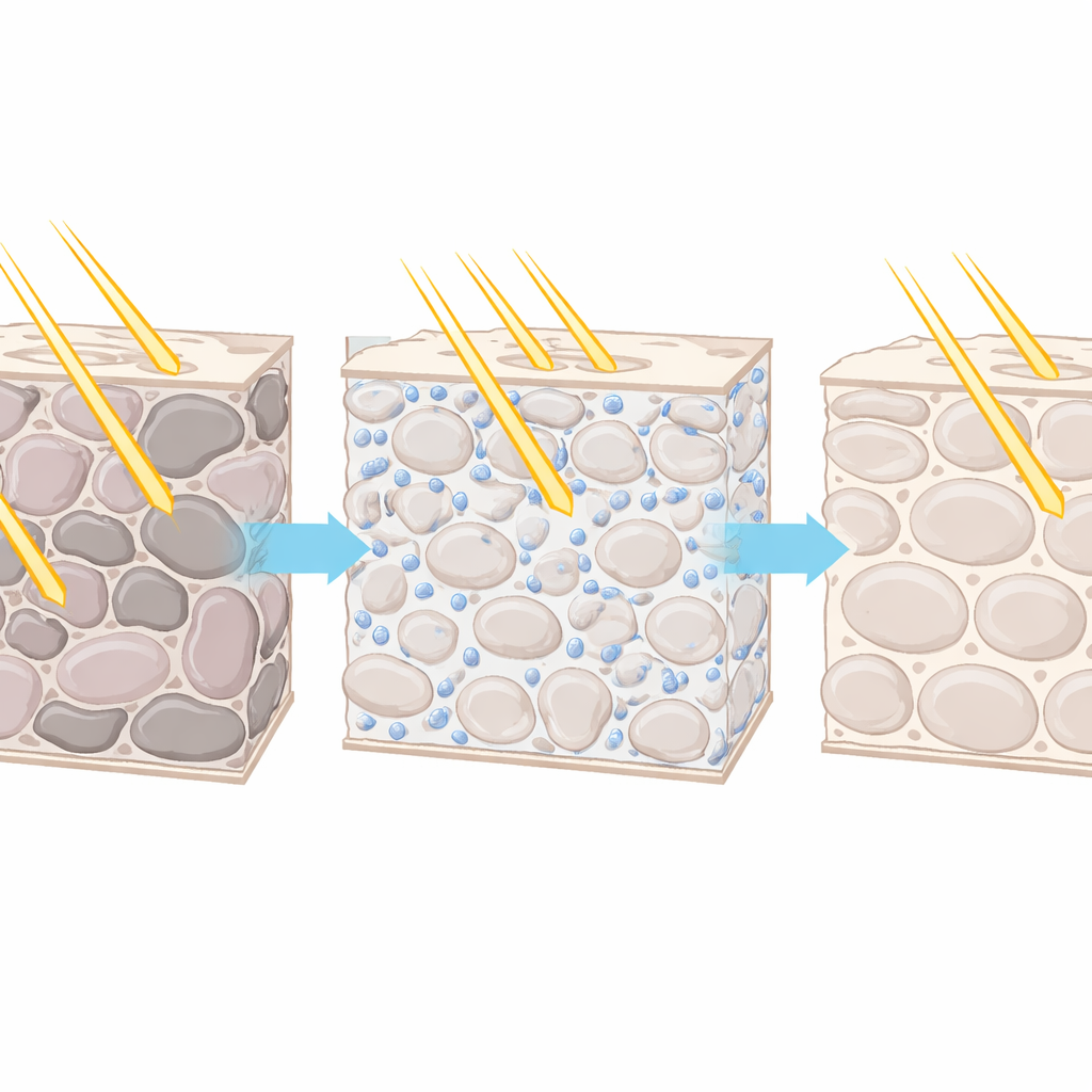
Living tissues are not naturally transparent because they are made of many tiny parts—cell membranes, organelles, fibers—with slightly different optical properties. As light passes through, it bends and scatters at each boundary, so conventional microscopes can only see a few hundred micrometers deep in the mouse brain. Existing “tissue clearing” cocktails solve this for fixed, dead samples by dissolving lipids or soaking tissues in thick, high-index liquids. But these mixtures are far too harsh for living cells: they pull water out of tissues, scramble salt balance, or seep into cells and disrupt normal activity, making them unsuitable for studying natural brain signals or organ function.
A Gentle Recipe Built Around Blood Protein
The authors reasoned that if they could match the optical density of the fluid outside cells to that of the watery cytosol inside them, light would scatter less, and tissues would appear clearer, even while alive. They screened many chemicals, including familiar ones like glycerol and a variety of medical contrast agents and polymers. Membrane-permeable molecules cleared cells but wiped out normal calcium responses, a basic sign of cell health. Long, chain-like polymers raised the salt concentration to damaging levels. A key insight was that compact, spherical macromolecules, especially the blood protein bovine serum albumin (BSA), could raise the refractive index of the medium with very little change in osmolarity. By carefully tuning BSA concentration and ions such as calcium and magnesium, they arrived at SeeDB-Live, a solution whose optical index closely matches that of the cell interior while keeping salt and water balance essentially physiological.
Making Organoids, Spheroids, and Slices Transparent
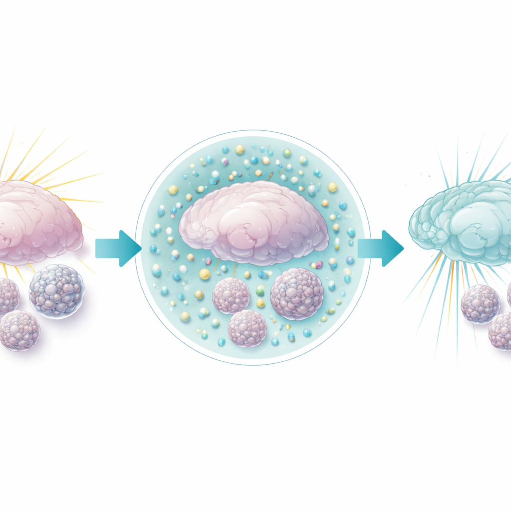
With SeeDB-Live in hand, the team tested it on increasingly complex living structures. Clusters of cancer-derived HeLa cells grown as spheroids, and miniature gut and brain organoids, rapidly became more transparent without swelling or shrinking. Under standard media, fluorescent signals faded after about 100 micrometers of depth; in SeeDB-Live, signals remained bright more than twice as deep. Importantly, these structures continued to grow and respond to stimuli such as high potassium, indicating that basic physiology remained intact. Acute brain slices from mice, bathed in SeeDB-Live, similarly cleared within about half an hour. Two-photon and confocal microscopes could then resolve neurons, dendrites, and fine structures much deeper in the cortex and hippocampus than before, and “shadow imaging” of all cells in a slice became feasible throughout the thickness, not just at the damaged surface.
Preserving Brain Function While Seeing More
Because even subtle changes in salt balance can alter nerve activity, the authors carried out detailed tests in mouse brain slices. Patch-clamp recordings from specific cortical neurons showed that resting voltage, firing thresholds and spike patterns under SeeDB-Live were very similar to those in standard artificial cerebrospinal fluid, with only modest parameter shifts. Calcium imaging in olfactory bulb slices revealed that spontaneous and evoked activity patterns were preserved in frequency and amplitude, even as signals became brighter at deeper layers. In contrast, other candidate clearing agents such as glycerol and iodixanol either suppressed spontaneous firing or slowed growth over days, highlighting the relative gentleness of the BSA-based solution.
Seeing into the Live Mouse Brain
The researchers then moved to living mice. After creating a small window in the skull and gently opening the protective membrane, they allowed SeeDB-Live to wash over the brain surface. Labeled albumin penetrated roughly half a millimeter into the cortex, and two-photon imaging of fluorescent neurons showed up to threefold brighter signals from deep cell bodies. Fine structures, such as dendritic spines hundreds of micrometers below the surface, became sharply visible. Tests of movement, feeding, and motor coordination before and after treatment showed no detectable behavioral side effects, and microscopic inspection of brain tissue revealed no surge in inflammation or cell death, even after repeated treatments over months.
Expanding What We Can Measure with Light
With better clarity, the team could push beyond calcium imaging to even more demanding readouts. In both brain slices and live animals, they used fast, camera-based epifluorescence to record voltage changes from genetically labeled neurons, including action potentials traveling along dendrites and synchronized firing across related cells in the olfactory bulb. These measurements, previously limited by scattering and low signal-to-noise, now became practical at higher speeds and over larger fields of view. Because SeeDB-Live is transient, the brain gradually returns to its original state as albumin is washed away, yet the process can be repeated through a simple plastic-covered cranial window for chronic studies.
What This Means for Future Brain and Organ Research
In essence, SeeDB-Live offers a way to temporarily “de-fog” living mammalian tissues without noticeably disturbing how their cells work. By matching the optical properties of the fluid between cells to that of their interior, the solution allows light to penetrate deeper with less distortion, enabling clearer images of structure and activity in everything from cell clusters to intact mouse brains. This advance opens the door to more routine deep imaging using standard microscopes, and to ambitious experiments that track fast electrical signals across many neurons in three dimensions, bringing us closer to watching whole circuits and organ-scale processes unfold in real time.
Citation: Inagaki, S., Nakagawa-Tamagawa, N., Huynh, N.Z. et al. Isotonic and minimally invasive optical clearing media for live cell imaging ex vivo and in vivo. Nat Methods 23, 839–853 (2026). https://doi.org/10.1038/s41592-026-03023-y
Keywords: tissue clearing, live imaging, two-photon microscopy, neural activity, organoids