Clear Sky Science · en
Mechanism of co-transcriptional cap snatching by influenza polymerase
How Flu Viruses Steal Our Cells’ Message Tags
Every winter, influenza viruses race through human populations, yet their success depends on a surprisingly delicate heist that unfolds deep inside our cells’ nuclei. To build their own genetic messages, flu viruses cannot make the protective “cap” that cells normally add to the front end of their RNAs. Instead, they steal this cap directly from our freshly made RNAs in a maneuver called “cap snatching.” This study reveals, in molecular detail, how the flu virus latches onto the human transcription machinery and cuts away these caps to power its own replication.
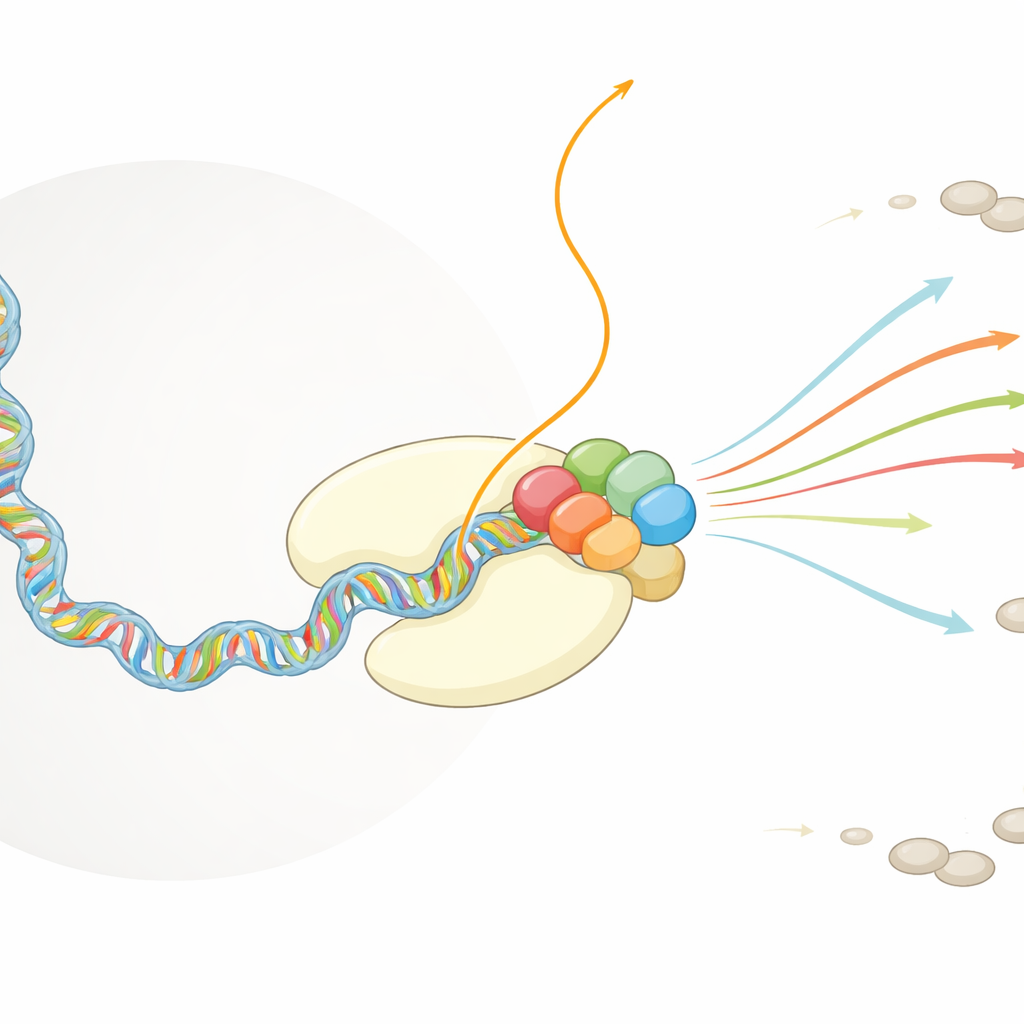
The Cell’s Copy Machine and the Viral Hitchhiker
Human cells rely on a massive protein assembly, RNA polymerase II, to read DNA and produce RNA copies that will later be turned into proteins. As soon as an RNA emerges from this machine, other cell factors quickly decorate its front end with a protective cap structure that helps stabilize the molecule and ensures it will be exported from the nucleus and translated. Influenza virus operates in the same nuclear space. Its own polymerase complex, called FluPol, cannot manufacture caps but depends absolutely on capped RNA fragments to start copying the viral genome into messenger RNAs. Earlier work showed that FluPol binds to RNA polymerase II, but how this interaction positions FluPol to grab and cut host RNA was unknown.
Capturing the Flu–Host Partnership in Action
To dissect this process, the researchers reconstituted a key portion of the human transcription apparatus in a test tube: RNA polymerase II engaged on DNA, producing a capped RNA, and accompanied by an elongation factor called DSIF that grips the emerging RNA. They then added FluPol and used cryogenic electron microscopy to visualize the combined complex at near‑atomic resolution, both before and after the viral cut takes place. Biochemical assays in parallel measured how efficiently FluPol could cleave host RNA under different conditions, such as with or without chemical marks on the tail of RNA polymerase II or with DSIF present.
Where and How the Viral Cut Is Made
The structures show FluPol nestling against the side of RNA polymerase II exactly where the new RNA exits. One part of FluPol recognizes the modified tail of polymerase II when it carries specific phosphate groups—a hallmark of very early transcription. Another part, the PB2 “cap‑binding” region, wedges into a groove on the surface of polymerase II right below the RNA exit channel and clamps onto the cap at the RNA’s front end. Meanwhile, the PA “endonuclease” region of FluPol contacts DSIF, which has rotated from its usual position to make room and help guide the RNA toward the viral cutting site. Together, these contacts stabilize the complex so that the endonuclease can slice the host RNA about 10–15 building blocks from the cap, generating a short capped fragment the virus can use.
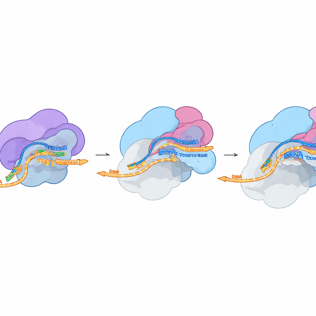
From Stolen Fragment to Viral Message
In a second structural snapshot taken under conditions that allow cutting, the researchers found that FluPol remains attached to the host machinery even after the RNA is cleaved. The cap stays firmly held in the PB2 pocket, but the freshly exposed end of the stolen fragment swings toward the active center of FluPol, where copying of viral RNA begins. The overall shape of FluPol in this state closely matches a previously described “pre‑initiation” form that is poised to start synthesis. This suggests that only minimal shape changes are needed to transition from cap snatching to full‑blown viral transcription. Cell‑based experiments, in which individual contact points between FluPol, DSIF and polymerase II were mutated, showed that disrupting these tiny interfaces sharply reduces viral polymerase activity and, in some cases, overall virus fitness.
Why These Findings Matter
This work reveals that influenza does not grab caps from just any RNA; it targets a very early stage of human transcription where polymerase II, its modified tail, DSIF, and a newly completed cap all come together. FluPol docks onto this composite platform, uses DSIF to present the RNA to its cutting site, trims off a capped fragment, and then feeds that fragment directly into its own copying machinery to launch viral mRNA production. For a lay observer, the key takeaway is that flu’s success hinges on a precise physical embrace between viral and host proteins. Although such small, flat contact surfaces are challenging drug targets, these atomic‑level maps now pinpoint exactly where a future antiviral could wedge in to disrupt the heist and blunt influenza infection.
Citation: Rotsch, A.H., Li, D., Dupont, M. et al. Mechanism of co-transcriptional cap snatching by influenza polymerase. Nature 652, 1281–1288 (2026). https://doi.org/10.1038/s41586-026-10189-0
Keywords: influenza virus, cap snatching, RNA polymerase II, viral transcription, cryo-EM structure