Clear Sky Science · en
Neuroimaging evidence of microstructural alteration in Parkinson’s disease with subjective cognitive decline
A Hidden Early Warning Sign in Parkinson’s
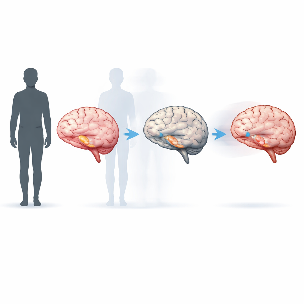
Many people think of Parkinson’s disease mainly as a movement disorder, marked by tremor and stiffness. Yet for a large share of patients, problems with memory, attention, and thinking can be just as disabling. This study asks a pressing question for patients and families: when someone with Parkinson’s starts to notice subtle lapses in memory or focus—before standard tests show anything wrong—does that “gut feeling” reflect real, measurable changes inside the brain?
From Everyday Slips to Measurable Brain Changes
The researchers focused on a group called “subjective cognitive decline,” or SCD. These are people with Parkinson’s who report frequent forgetfulness or trouble concentrating, but who still score within the normal range on routine cognitive tests. The team compared four groups: people with Parkinson’s and normal cognition, people with Parkinson’s plus SCD, people with Parkinson’s and mild cognitive impairment, and healthy adults without Parkinson’s. All participants underwent detailed cognitive testing and advanced MRI scans that can reveal tiny changes in brain wiring and memory-related structures.
Looking Deep into Brain Wiring
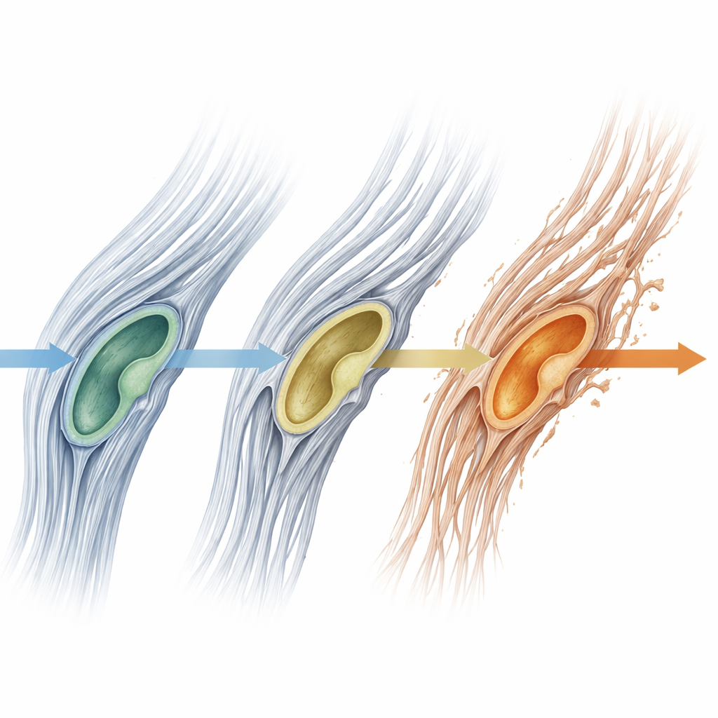
To examine the brain’s communication highways, the scientists used a diffusion MRI technique that tracks how water moves along white matter fibers. One measure, called the peak width of skeletonized mean diffusivity (PSMD), summarizes overall white matter injury; another, fractional anisotropy (FA), captures the health of specific fiber bundles. They also used high-resolution scans to break the hippocampus—a key memory hub—into smaller subregions and measure their sizes. These approaches allowed the team to detect very subtle brain changes that would be invisible on standard clinical images.
Early Damage Appears Before Standard Tests Fail
The results showed a clear pattern across the four groups. White matter injury, as measured by PSMD, was lowest in healthy volunteers and in people with Parkinson’s but no cognitive complaints, higher in those with Parkinson’s plus SCD, and highest in those with mild cognitive impairment. Higher PSMD values went hand-in-hand with worse performance on global thinking tests, especially memory and attention. When the researchers zoomed in on specific nerve fiber tracts, they found widespread damage in patients with mild cognitive impairment, while those with SCD showed only a small, borderline change in a major fiber bundle connecting the two halves of the brain. This suggests that broad, diffuse damage to white matter may build up before most local changes are obvious.
Memory Center Shrinkage Starts Early
Even more striking, people with Parkinson’s and SCD already showed shrinkage in certain subregions of the hippocampus, including areas deeply involved in forming new memories and linking memory with emotion. These same subregions were also smaller in patients with mild cognitive impairment, but overall hippocampal size did not differ between groups. In other words, the earliest changes were only visible when the hippocampus was dissected into fine-grained parts. One particular transition zone between the hippocampus and the amygdala was closely tied to both memory scores and self-reported cognitive complaints, hinting at a biological bridge between mood, emotion, and memory worries in Parkinson’s.
What This Means for Patients and Care
This study suggests that when a person with Parkinson’s reports frequent memory lapses or trouble focusing, those concerns should not be dismissed simply because basic tests look normal. Subtle but meaningful changes in brain wiring and in key memory regions already appear to be under way at this stage. The PSMD measure, which captures global white matter injury, stood out as a particularly sensitive marker for distinguishing stages of cognitive change in Parkinson’s. While more and larger long-term studies are needed, especially ones that track individuals over time, these findings point toward new brain-based tools that could one day help doctors identify higher-risk patients earlier and tailor monitoring, counseling, and treatment before everyday thinking problems become harder to reverse.
Citation: Chen, K., Zhang, R., Ji, Y. et al. Neuroimaging evidence of microstructural alteration in Parkinson’s disease with subjective cognitive decline. npj Parkinsons Dis. 12, 105 (2026). https://doi.org/10.1038/s41531-026-01313-y
Keywords: Parkinson’s disease, subjective cognitive decline, white matter changes, hippocampal atrophy, brain MRI