Clear Sky Science · en
Thyrotropin-releasing hormone neurons of different hypothalamic nuclei increase energy expenditure
Why brain cells that burn calories matter
Most people think of metabolism as something controlled by hormones from glands like the thyroid. This study looks deeper, into tiny clusters of brain cells that release a messenger called thyrotropin‑releasing hormone (TRH) in mice. The researchers show that different groups of these cells act like separate “energy hubs,” each tuning how much heat the body makes, how much it moves, and how much it eats. Understanding these circuits could point to new ways to fight obesity and diabetes by nudging the body to waste more calories as heat instead of storing them as fat.
Different brain hubs, one common fuel goal
The team focused on several regions in the hypothalamus, a key control center deep in the brain, and on a brainstem area. They asked whether TRH‑producing neurons in each region help control the body’s energy use. Using viruses as tracers, they mapped connections between these neurons and brown adipose tissue, a special type of fat that burns energy to produce heat. They then used a chemogenetic switch—an engineered receptor activated by a harmless designer drug—to turn selected TRH neuron groups on and off in living mice while measuring body temperature, brown fat activity, movement, and food intake.
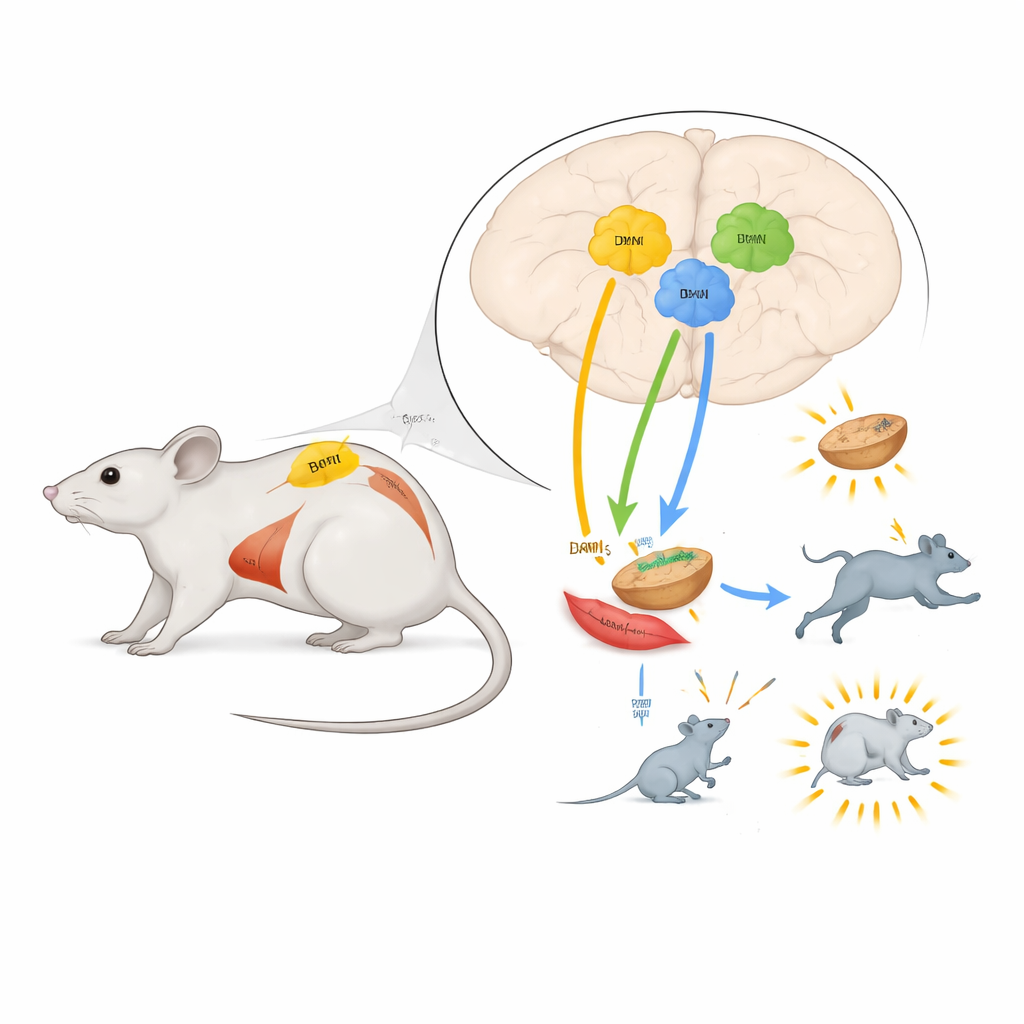
Brown fat burners in two key regions
In the paraventricular nucleus (PVN) and the dorsomedial hypothalamus (DMH), activating TRH neurons sharply increased how many calories the mice burned and raised their core body temperature. Infrared imaging showed that brown fat patches between the shoulder blades warmed up, and molecular tests confirmed that enzymes involved in fat breakdown inside brown fat were switched on. Blocking a specific type of adrenaline receptor in fat cells prevented this heating, indicating that these brain cells drive brown fat through the sympathetic nerves, the same system that prepares the body for “fight or flight.” These changes in energy use and temperature persisted even when the animals were not allowed to eat, showing that they were not just a side effect of increased food intake.
A movement hub that protects against the cold
A different picture emerged in the medial preoptic area (MPA), a region long known to sense body temperature. Turning on TRH neurons here also raised energy use and body temperature, but brown fat stayed relatively quiet. Instead, the mice became more active: they moved around more in their cages, suggesting that muscle work and general arousal were supplying much of the extra heat. When the researchers chronically silenced these MPA TRH neurons and then exposed mice to a sudden drop in room temperature, the animals’ body temperature fell more steeply and they failed to boost their energy output. This shows that MPA TRH neurons are essential for mounting a proper cold‑defense response, likely by driving behavior and muscle activity rather than directly switching on brown fat.
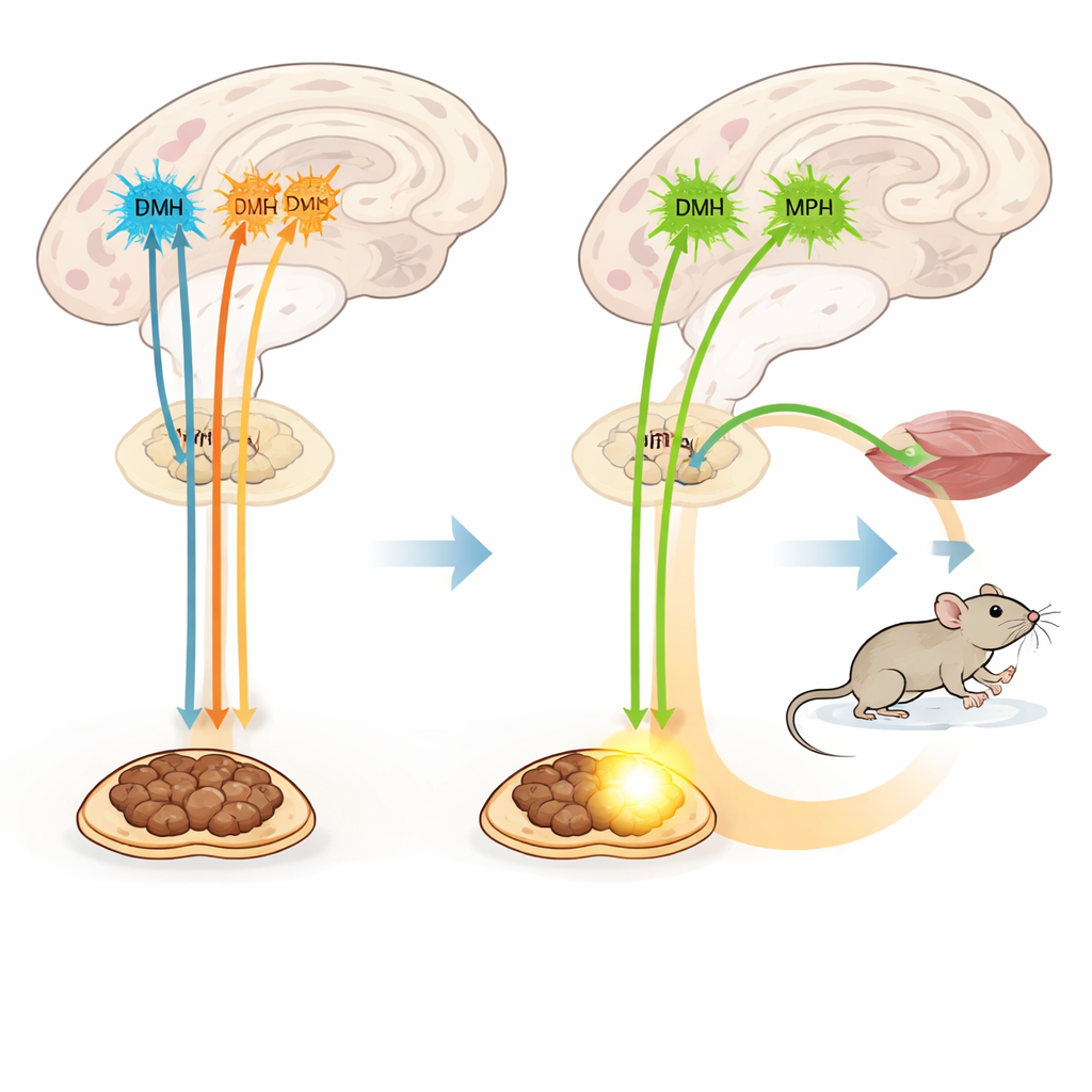
Not all TRH cells shape metabolism
The scientists also examined TRH neurons in a brainstem region called the rostral raphe pallidus, which had been suspected to help control brown fat. Surprisingly, activating these cells had little effect on energy use, movement, brown fat temperature, or eating. This suggests that simply being connected to brown fat is not enough; only some TRH‑positive circuits actually change how much energy the body spends.
Beyond classic thyroid hormones
TRH is best known for triggering release of thyroid‑stimulating hormone and thyroid hormones, which broadly raise metabolism. Here, only the PVN TRH neurons activated this hormone chain. Yet the rapid boosts in brown fat heat and energy use driven by PVN and DMH TRH neurons did not depend on the main TRH receptor that controls thyroid hormone release. Even when this receptor was genetically deleted, turning on PVN TRH neurons still made brown fat hotter and body temperature rise. A TRH‑like drug, by contrast, required that receptor to increase energy use. This split shows that the same chemical messenger supports at least two systems: a slower, whole‑body hormonal one and a faster, nerve‑based one routed through specific TRH neuron groups.
What this means for human health
In everyday terms, the study reveals that several small sets of TRH‑releasing brain cells work together like specialized thermostats and fuel valves. Those in two hypothalamic hubs directly push brown fat to burn calories as heat, while those in a neighboring region boost movement and help the body cope with cold. All three hypothalamic groups also briefly increase eating, likely by exciting hunger‑related circuits. Because these effects can be separated from classic thyroid hormones, targeting the right TRH pathways or their nerve connections might one day allow doctors to dial up energy expenditure without triggering the wide‑ranging side effects of excess thyroid hormone.
Citation: Constantinescu, A., Chandrasekar, A., Kleindienst, L. et al. Thyrotropin-releasing hormone neurons of different hypothalamic nuclei increase energy expenditure. Nat Commun 17, 3499 (2026). https://doi.org/10.1038/s41467-026-71617-3
Keywords: brown fat thermogenesis, hypothalamus, thyrotropin-releasing hormone, energy expenditure, cold tolerance