Clear Sky Science · en
iCLAP: an innovative method for integrable co-detection of low-abundance antigens with high-plex immunostaining
Seeing Hard-to-Spot Clues in Everyday Tissues
Doctors and scientists often rely on thin slices of preserved tissue, stored for years in hospital archives, to understand how diseases like cancer or diabetes develop. But some of the most important warning signs in these samples—proteins that appear only in tiny amounts—are nearly invisible with current imaging tools. This study introduces a new method, called iCLAP, that turns these faint molecular whispers into clear signals, all while using the same routine tissue blocks already found in pathology labs.
Making Faint Signals Shine
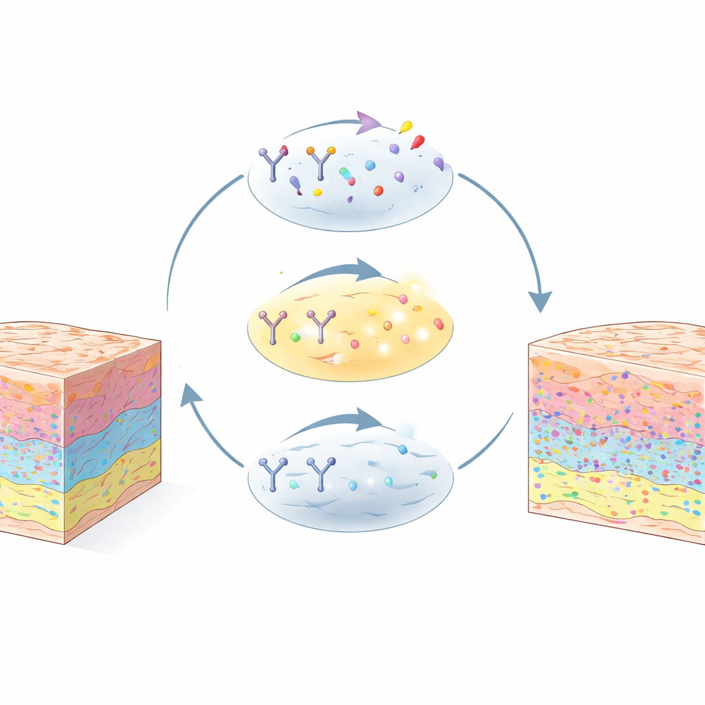
Most advanced imaging methods can look at many proteins at once, but they work best for those that are present in high amounts. Key regulators of aging, immune escape, and cancer behavior are often rare and get missed. iCLAP (Integrable Co-detection of Low-Abundance Proteins) solves this problem by building on a chemistry trick known as signal amplification. The method first uses an enzyme-driven reaction to stack many fluorescent tags near each target protein, greatly boosting the light it gives off. Then, a carefully tuned bleaching step removes that strong signal without harming the tissue or the underlying proteins. This allows the same tissue slice to be stained, imaged, erased, and reused across many cycles.
Working With the Tissues Clinics Already Have
Crucially, iCLAP is designed for formalin-fixed, paraffin-embedded (FFPE) tissue—the standard way hospitals preserve biopsies and surgical samples. The authors show that repeated amplification and bleaching cycles cause only minimal tissue loss, comparable to existing multiplex imaging methods. After the low-abundance proteins are imaged with iCLAP, the same section can be stained again with more conventional approaches to reveal abundant structural or cell-type markers. iCLAP can be combined with several popular high-plex platforms, including CyCIF, CODEX, and imaging mass cytometry, enabling maps of more than 40 different proteins in a single tissue slice.
Tracking Cellular Aging in the Pancreas
To demonstrate what this added sensitivity makes possible, the team focused on cellular senescence—an altered state in which cells stop dividing and often change their behavior. Senescent cells are thought to influence aging, diabetes, and cancer, but the proteins that mark this state can be very scarce in human tissues. Using iCLAP, the researchers could clearly detect multiple senescence markers, including P16, P21, P53, 53BP1, HMGB1, and Lamin B1, in stored sections of human pancreas. When they compared iCLAP-based amplification with standard fluorescent staining, many markers that were almost invisible before became sharply defined. This allowed them to measure marker levels in tens of thousands of individual cells and to group cells into distinct subpopulations based on their senescence profiles.
Linking Aging Signals to Tissue Structure and Function
With this more detailed view, the scientists mapped where senescence-related proteins appeared within the pancreas. They found that different markers tended to dominate in different compartments: some were enriched within the hormone-producing islets, others in the enzyme-producing acinar regions, and others in ductal structures. Within islets, cells carrying high levels of the marker P16 were more common in larger and older islets and were associated with shifts in the balance of insulin- and glucagon-producing cells. At the single-cell level, however, most cells expressed only one senescence marker strongly, suggesting that the “aging” state in human tissues is more diverse and fragmented than the broad, multi-marker patterns often seen in lab-grown cells.
A Versatile Window Into Subtle Disease Signals
Finally, the team applied iCLAP to a range of normal and tumor tissues from organs such as breast, liver, cervix, ovary, and skin. Across these samples, tumors showed much stronger senescence-marker signals than nearby healthy tissue, underscoring the method’s potential for cancer research. By making low-level proteins visible while preserving the ability to look at dozens of markers at once, iCLAP turns archived clinical samples into rich, high-dimensional maps of cell states and neighborhoods. For non-specialists, the key message is that many crucial disease signals have been hiding in plain sight in existing tissue collections—and this new approach offers a practical way to reveal them.
Citation: Wu, F., Zheng, S., Chen, Y. et al. iCLAP: an innovative method for integrable co-detection of low-abundance antigens with high-plex immunostaining. Nat Commun 17, 3104 (2026). https://doi.org/10.1038/s41467-026-69752-y
Keywords: spatial proteomics, cellular senescence, multiplex imaging, FFPE tissue, pancreatic islets