Clear Sky Science · en
Structural and functional mechanisms underlying activation gate dynamics and IFM motif accessibility in human Nav1.5
Why tiny heart gates matter
The electrical beat of every heartbeat depends on microscopic gates in heart cells that briefly open to let sodium ions rush in. These gates are proteins called sodium channels, and when they misfire, the result can be dangerous heart rhythm disorders. This study focuses on the main cardiac sodium channel, known as Nav1.5, and reveals a previously unseen in‑between state of its gate, plus a hidden ion pocket next to a key “latch” that turns the channel off. Understanding this machinery in such detail could guide better drugs for arrhythmias that act with greater precision and fewer side effects.
A closer look at the heart’s sodium door
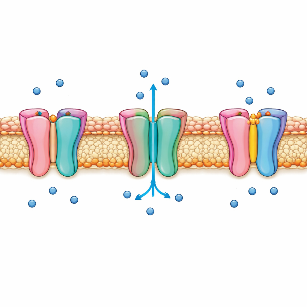
Nav1.5 sits in the heart cell membrane and opens for only a few thousandths of a second to generate the sharp rise of the electrical signal that triggers contraction. Almost immediately, it switches itself off in a process called fast inactivation, preventing excessive sodium entry. For decades, a short three‑amino‑acid segment, nicknamed the IFM motif, has been thought to swing into place like a plug to stop the flow. Yet many details of how the main gate at the bottom of the channel moves, and how the IFM motif actually controls this motion, have remained unclear. These gaps have limited efforts to design medicines that target the channel without disturbing its finely tuned timing.
Capturing an in‑between open state
Using high‑resolution cryo‑electron microscopy, the authors captured the full‑length human Nav1.5 channel in what they call an intermediate open state. In this configuration, the four inner helices that form the activation gate are spread just wide enough to allow hydrated sodium ions to pass, but not as wide as in fully open structures described previously. Molecular simulations under applied voltage showed that water and sodium ions can cross the pore, though only at higher voltages than in a fully open channel, confirming that this state conducts but sits between the usual open and inactivated forms. A detergent molecule lodged at the gate helps hold this delicate arrangement, illustrating how small changes in shape can finely tune whether the channel passes ions or remains shut.
How side contacts help control the gate
The team also resolved an interaction surface between the channel’s N‑terminal domain, which hangs inside the cell, and one of the gate‑forming helices known as S6 in domain I. Specific charged amino acids in these regions form salt bridges that act like tiny latches helping to stabilize the open gate. When the researchers altered these residues, the overall shape of the electrical currents changed: in particular, some mutations slowed how quickly the channel turned off after opening. Additional analyses revealed that the S6 helix can rock inward and outward, narrowing or widening the gate. Different patterns of contact with the N‑terminal domain matched these shifts, tying the microscopic motion of a single helix to the channel’s ability to switch between open and more closed, inactivated‑like shapes.
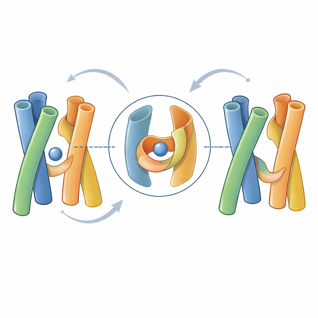
A hidden pocket beside the inactivation latch
Perhaps the most striking discovery lies beside the IFM motif itself. In the intermediate open structure, the IFM segment retains its characteristic U‑shaped pose snugly docked in a hydrophobic pocket, rather than swinging completely away as some models propose. Next to it, however, the authors identified a second pocket lined with negatively charged residues that holds a sodium ion. Simulations and experiments suggest that other positively charged ions can also occupy this niche. Mutations that changed the charge or chemistry of the pocket altered how easily the channel inactivated and how the IFM motif behaved, indicating that ion binding here loosens or tightens the IFM latch without requiring it to fully leave its cradle. This reinterpretation helps explain past experiments where reactive ions could label engineered cysteines near the IFM motif even though the motif appeared to remain buried.
What this means for heart rhythm and future drugs
Together, these findings update the classic “door‑and‑wedge” picture of sodium channel inactivation. Rather than a simple plug being pulled in and out, Nav1.5 appears to use a coordinated dance: a gate that can sit in an intermediate open position, side contacts that steady or relax this configuration, and a nearby ion‑tuned pocket that adjusts how firmly the IFM motif holds the gate shut. By revealing this alternative ion access pathway and its impact on gating, the work offers a refined structural blueprint for how cardiac sodium channels fail in disease and points to new, more selective targets for anti‑arrhythmic therapies.
Citation: Biswas, R., López-Serrano, A.L., Purohit, A. et al. Structural and functional mechanisms underlying activation gate dynamics and IFM motif accessibility in human Nav1.5. Nat Commun 17, 2820 (2026). https://doi.org/10.1038/s41467-026-69672-x
Keywords: cardiac sodium channel, Nav1.5, fast inactivation, cryo-EM, cardiac arrhythmia