Clear Sky Science · en
Nucleoporin TPR integrates MAPK signaling with mitogen-induced transcriptional programs
How Cells Decide When to Grow
Our cells constantly receive go-or-stop signals telling them when to divide, rest, or react to their surroundings. Many of these commands travel through a well-known chain of proteins called the MAPK pathway, which is often overactive in cancer. This study uncovers an unexpected player at the gateway to the cell’s nucleus—a large scaffold protein named TPR—that helps fine‑tune how growth signals are translated into bursts of gene activity. Understanding this extra layer of control may open new ways to read, and eventually adjust, cancer‑related signaling in patients.
A Gatekeeper at the Edge of the Nucleus
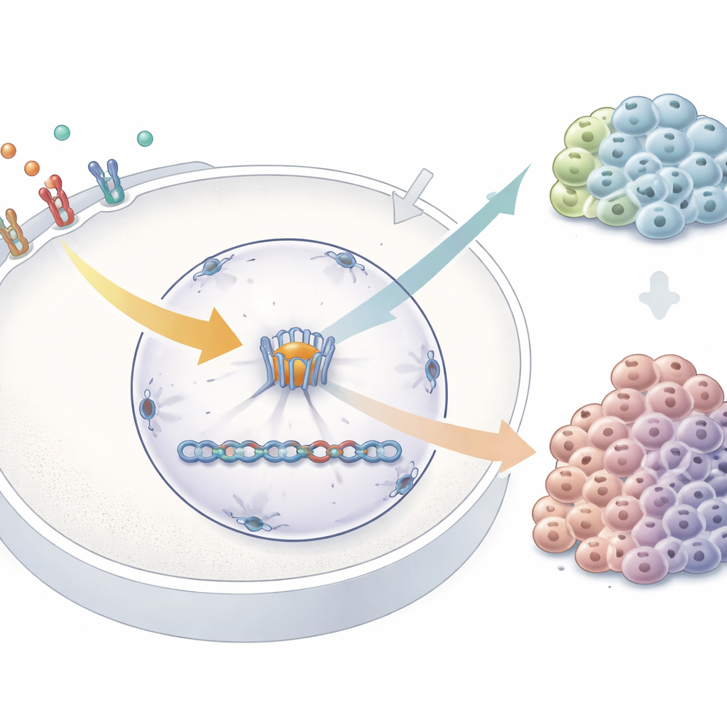
Every cell nucleus is studded with thousands of nuclear pores—massive channels that let molecules move in and out. TPR is a long structural protein attached to the inner side of these pores. It was known to help with tasks such as exporting RNA and maintaining genome stability, but its role in growth control was unclear, even though its levels are abnormal in several cancers. The authors asked whether TPR is not just a passive part of the pore but also a responsive element that links incoming growth cues to changes in gene activity deep inside the nucleus.
Listening to Growth Signals at the Nuclear Pore
To probe this idea, the researchers reduced TPR levels in human cancer cells and then stimulated them with epidermal growth factor, a classic trigger of the MAPK signaling chain. Using RNA sequencing, they compared gene activity in normal versus TPR‑depleted cells, both at rest and after stimulation. They found that hundreds of genes changed their behavior when TPR was missing, including many tied to the MAPK pathway itself. Some genes linked to growth promotion and feedback control were already altered in unstimulated cells, while others changed only after the growth signal arrived. Notably, an immediate‑early gene called FOS—one of the fastest responders to MAPK activation—was turned on more strongly when TPR was depleted, and this overshoot depended on active growth‑factor signaling. Functionally, cells lacking TPR responded abnormally to prolonged stimulation, showing a shifted pattern of cell‑cycle progression.
A Chemical Switch on TPR
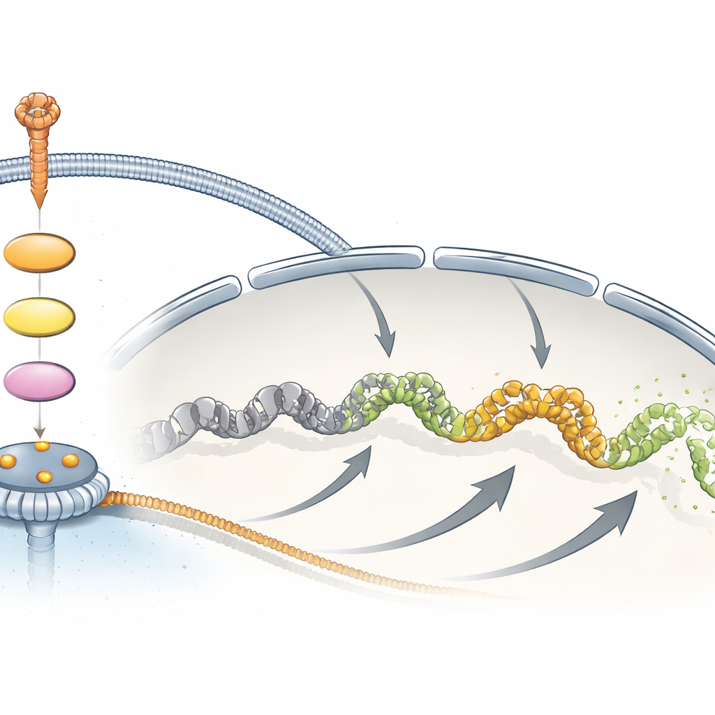
The team then looked for a physical connection between MAPK signaling and TPR. Earlier large‑scale protein surveys had hinted that a specific site on TPR, an amino acid called Ser2155, becomes phosphorylated—that is, chemically tagged—after growth‑factor treatment. The authors created a highly specific antibody that recognizes TPR only when this site is phosphorylated, allowing them to track this modification over time and across systems. In multiple human cell types, growth‑factor stimulation rapidly and transiently increased phosphorylation at Ser2155, closely tracking the activation of ERK, a key MAPK enzyme. Blocking the upstream growth‑factor receptor, or the MEK‑ERK steps of the pathway, sharply reduced this phosphorylation. Removing ERK1 and ERK2 together nearly abolished it, while eliminating TPR itself did not change ERK activity, placing TPR clearly downstream of the MAPK cascade rather than as a core signaling component.
From Mice to Human Tumors
To test whether this connection matters beyond cell lines, the researchers generated mice with only one working copy of the Tpr gene; complete loss proved lethal during development. In their spleens—a tissue where immune cells activate the MAPK pathway through their antigen receptors rather than through the growth‑factor receptor—the overall pattern of gene activity shifted, again enriching for MAPK‑related genes and dampening several natural brakes on the pathway. When splenocytes from these mice were stimulated through their T‑cell receptors, they produced more Fos messenger RNA than cells from normal animals, mirroring the human cell‑culture results. The team then examined human tumor samples. In most serous ovarian cancers, and in a large subset of triple‑negative breast cancers, staining for phosphorylated TPR at Ser2155 was common, though breast tumors showed much more case‑to‑case variability, consistent with their mixed patterns of pathway activation.
Why This Matters for Cancer and Therapy
Taken together, the findings paint TPR as a responsive nuclear factor that sits at the nuclear pore and is chemically tuned by the MAPK pathway. Rather than turning the pathway on or off, TPR helps shape the amplitude and quality of the transcriptional burst that follows growth or immune stimulation, particularly for rapidly induced genes like FOS. Because many cancers rely on persistent MAPK activity, the presence and phosphorylation status of TPR may serve as a useful readout of pathway engagement in tumors, and its mis‑regulation could contribute to distorted growth responses. This work broadens our view of nuclear pores from static gateways to active participants in how cells interpret mitogenic signals.
Citation: Liu, J., Zheng, Y., Xiong, Y. et al. Nucleoporin TPR integrates MAPK signaling with mitogen-induced transcriptional programs. Cell Death Dis 17, 400 (2026). https://doi.org/10.1038/s41419-026-08760-8
Keywords: nuclear pore, MAPK signaling, TPR protein, growth factor signaling, cancer transcription