Clear Sky Science · en
Structural brain differences associated with panic disorder: an ENIGMA-Anxiety Working Group mega-analysis of 4924 individuals worldwide
Why Sudden Waves of Fear Matter
Panic attacks can feel like drowning on dry land: a racing heart, shortness of breath, and an overwhelming sense that something terrible is about to happen. For people with panic disorder, these episodes strike again and again, often without warning, disrupting school, work, and relationships. This large international study asks a simple but important question: do the brains of people with panic disorder differ, on average, from those who have never experienced such attacks—and if so, where and how?
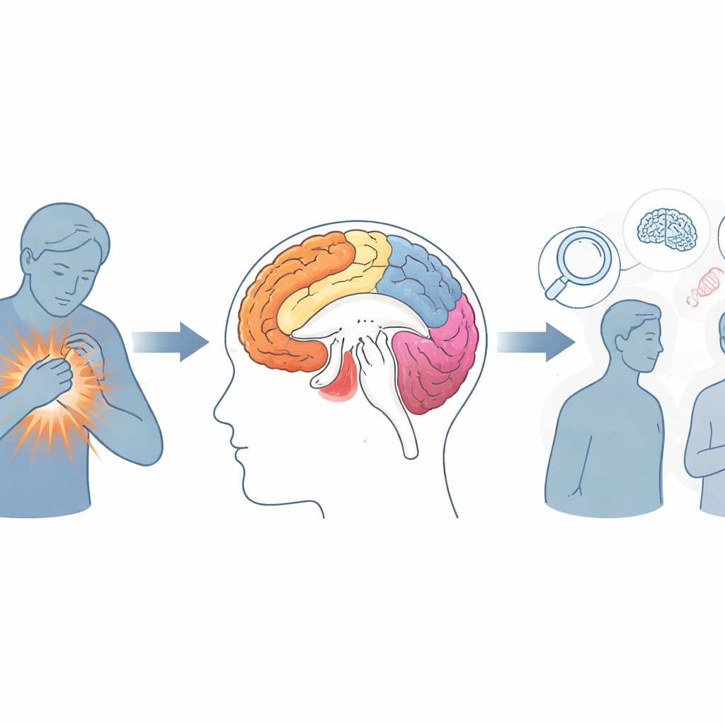
Looking Inside Thousands of Brains
To find out, researchers from 28 centers around the world pooled brain scans from nearly 5,000 participants aged 10 to 66. About one in four had been diagnosed with panic disorder, while the rest had no history of mental illness or psychiatric medication. All volunteers lay in an MRI scanner, which produces detailed pictures of brain structure. Using the same computerized methods at every site, the team measured how thick the brain’s outer sheet (the cortex) was, how much surface it covered, and how large key deep structures were, including the thalamus and the caudate nuclei. By harmonizing how data were collected and analyzed, the researchers could detect subtle differences that smaller, stand‑alone studies are usually too weak to pick up.
Fine Changes in the Brain’s Outer Shell
The cortex is a bit like the brain’s processing canvas: it helps us see, feel, remember, and regulate emotions. People with panic disorder showed slightly thinner cortex in several regions, including areas that help interpret sights, faces, and bodily sensations. These included parts of the temporal lobe and fusiform gyrus, which are involved in recognizing faces and reading body signals, as well as regions near the sensorimotor strip that help register and control bodily feelings. Surface area—how much space the cortex covers—was also a little smaller in a few left‑sided frontal, temporal, and parietal areas. Although these differences were small for any one person, they were consistent across many sites, hinting that long‑term vulnerability to panic may go hand‑in‑hand with slightly altered "wiring" for processing emotional and bodily cues.
Deep Hubs and Brain Fluid Spaces
Changes were not limited to the brain’s surface. In people with panic disorder, two deep relay hubs—the caudate nuclei and parts of the thalamus—were modestly smaller. These regions help link incoming information to habits, motivation, and defensive reactions. Their reduced size may contribute to the brain’s tendency to overreact to harmless bodily changes, such as a small shift in breathing or heart rate, and to form rigid fear responses around them. The study also found that individuals whose panic attacks began early in life (at or before age 21) had larger fluid‑filled spaces in the brain, called lateral ventricles, than those whose problems started later. This enlargement may reflect long‑term changes in surrounding brain tissue for early‑onset cases, although the study could not determine cause and effect.
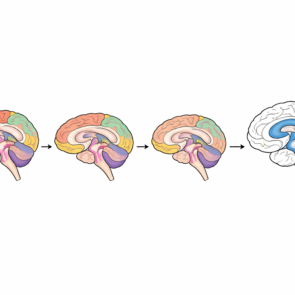
How Age Shapes the Picture
The team went a step further by asking whether age changed the relationship between panic disorder and brain structure. Instead of looking only at a straight line from youth to old age, they modeled a curved (non‑linear) pattern and discovered that differences in cortical thickness depended strongly on life stage. The clearest thinning in panic disorder appeared during adulthood, roughly between ages 25 and 55. In younger and older participants, the contrast between people with and without panic disorder was weaker and statistically uncertain. This suggests that the brain changes linked to panic disorder may emerge or become most apparent in midlife, potentially reflecting how the condition interacts with the brain’s natural development and aging.
What This Means for People Living With Panic
For individuals dealing with panic attacks, these findings do not mean that their brains are "damaged" or that change is impossible. Rather, the study shows that panic disorder is associated with subtle, widespread shifts in brain structure—especially in networks that process sensations, emotions, and bodily states—and that these shifts vary with age and the timing of onset. The differences are too small to diagnose or predict panic disorder from a single scan, but they provide a biological framework for why ordinary bodily sensations can feel so alarming and hard to control. As future research tracks people over time and combines structural scans with measures of brain activity and connectivity, these insights may guide more precise prevention and treatment strategies tailored to when in life panic first takes hold.
Citation: Han, L.K.M., Bruin, W.B., Bas-Hoogendam, J.M. et al. Structural brain differences associated with panic disorder: an ENIGMA-Anxiety Working Group mega-analysis of 4924 individuals worldwide. Mol Psychiatry 31, 2402–2417 (2026). https://doi.org/10.1038/s41380-025-03376-4
Keywords: panic disorder, brain structure, MRI, anxiety, neuroimaging