Clear Sky Science · en
Photoacoustic microscopy reveals deep angiogenic responses in 3D bioprinted tumor–vessel models
Why Looking Inside Tiny Tumors Matters
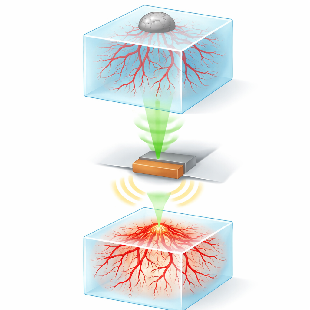
Cancer tumors do not grow in isolation; they build their own lifeline of blood vessels to bring in oxygen and nutrients. Many modern drugs try to cut off this lifeline, but testing how well such treatments work usually relies on animals or flat cell layers in dishes, each with serious drawbacks. This study presents a way to grow miniature three-dimensional tumor–blood vessel systems in the lab and then look deep inside them without cutting them open, using a sound-based imaging method called photoacoustic microscopy.
Building Mini Organs on a Chip
The researchers first created realistic, thumb‑sized cancer models using 3D bioprinting. They printed soft hydrogel blocks loaded with human blood vessel cells and supporting cells so that tiny capillary‑like networks would naturally form throughout the gel over several days. On top of these living vascular layers, they gently placed clusters of brain cancer cells (tumor spheroids). Over time, these tumors began to interact with the vessels below, encouraging new sprouts to grow toward them, much like tumors do in the body.
Listening to Light to See Deep Inside
Seeing what happens deep inside these cloudy, cell‑filled gels is difficult for standard microscopes because light gets scattered and quickly fades with depth. The team solved this by using high‑resolution photoacoustic microscopy. In this technique, short laser pulses shine into the tissue and are absorbed by certain molecules, causing tiny, rapid expansions that produce ultrasound waves. A small detector picks up these waves, and a computer reconstructs detailed three‑dimensional images. To make the vessels and cancer cells visible, the researchers used a common laboratory dye (MTT) that living cells convert into dark crystals, which strongly absorb the laser light and generate clear signals throughout the sample.
Watching Blood Vessels Grow in 3D
With this setup, the authors showed that their photoacoustic system could see significantly deeper into the bioprinted tissue than a standard confocal microscope—about 1.6 times deeper, reaching nearly a millimeter. They tracked how the vessel networks changed over several days: at first short, sparse branches appeared, then they elongated, intertwined, and formed longer, more complex paths. By digitally tracing each vessel in three dimensions, they quantified how average and maximum vessel lengths increased over time, confirming that the model faithfully captures the gradual buildup of tumor‑like blood supply.
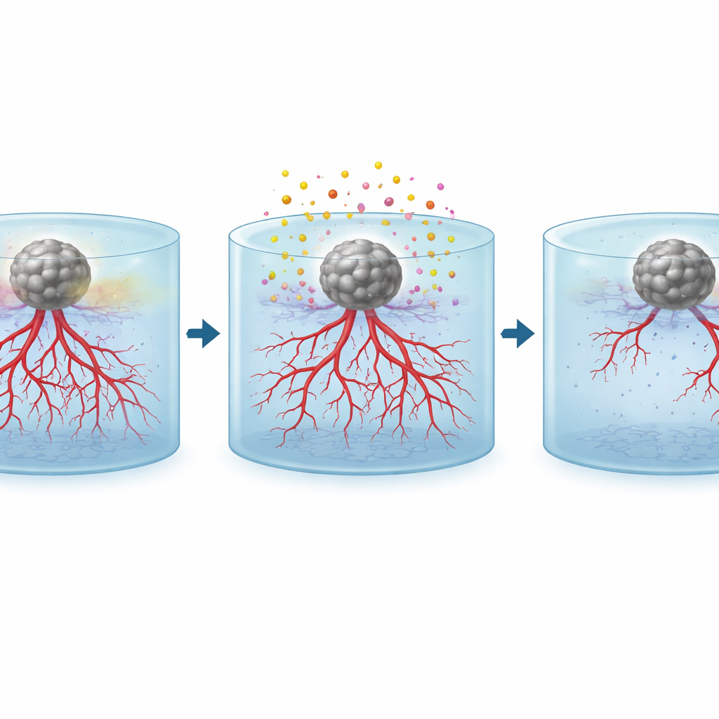
Testing Cancer Drugs in a Miniature Lab
The real power of the platform comes from using it to test treatments. After the tumor spheroids had begun to influence nearby vessels, the team applied two widely used anti‑cancer drugs: temozolomide, which mainly harms rapidly dividing tumor cells, and sunitinib, which directly blocks vessel‑growth signals. They tried each drug alone and in combination. Photoacoustic images clearly revealed that, compared with untreated samples full of dense, radiating vessel networks, drug‑treated samples had fewer, shorter, and more fragmented vessels, especially near the tumors. The combined treatment produced the strongest reduction in vessel density at all depths, demonstrating that this approach can measure how different therapies alter tumor‑driven vessel growth in 3D.
What This Means for Future Cancer Care
This work shows that it is possible to grow realistic, three‑dimensional tumor–vessel systems in the lab and then noninvasively “see” their internal blood vessel networks with sound generated by light. Because the method captures the entire volume, not just the surface, it can reveal where and how strongly a drug cuts off a tumor’s blood supply throughout the tissue. In the future, similar bioprinted constructs made from a patient’s own cells could help doctors compare treatment options before giving them, pointing the way toward faster, more ethical, and more personalized cancer therapy testing.
Citation: Jo, Y., Han, S., Kye, H. et al. Photoacoustic microscopy reveals deep angiogenic responses in 3D bioprinted tumor–vessel models. Microsyst Nanoeng 12, 129 (2026). https://doi.org/10.1038/s41378-026-01243-y
Keywords: tumor angiogenesis, photoacoustic microscopy, 3D bioprinting, cancer drug screening, vascular imaging