Clear Sky Science · en
Evaluation of lactic acid as a novel fixative for histological and neuroanatomical applications
Why safer tissue preservation matters
Whenever doctors or scientists study tissue under the microscope, they first need to “fix” it so cells do not decay. The traditional chemical for this job, formaldehyde, preserves structure very well but is toxic and linked to cancer risk for people who work with it every day. This study asks a simple but important question: could lactic acid, a common food preservative, offer a safer way to prepare brain samples while still keeping the fine details that pathologists and researchers rely on?
From food preservative to lab tool
Lactic acid is widely used to keep food, cosmetics, and household products free of harmful microbes. It is considered safe for consumers and has known antibacterial and tissue preserving effects. Because of this, the researchers explored whether lactic acid solutions could also stabilize brain tissue for standard microscopic staining, focusing on mouse brains as a model. They compared several lactic acid strengths with the current gold standard fixative, neutral buffered formalin, and with a salt solution that acts as a non fixing control.
Putting lactic acid to the test
The team used two common ways of fixing brain tissue. In immersion fixation, the removed brain is placed directly into the solution. In perfusion fixation, the solution is pumped through the animal’s blood vessels before the brain is removed, which usually gives more even preservation. The researchers tested lactic acid at different concentrations and for different lengths of time, then processed the samples in the usual way: embedding in wax, cutting very thin slices, and staining them with the standard hematoxylin and eosin dyes used in most pathology labs.
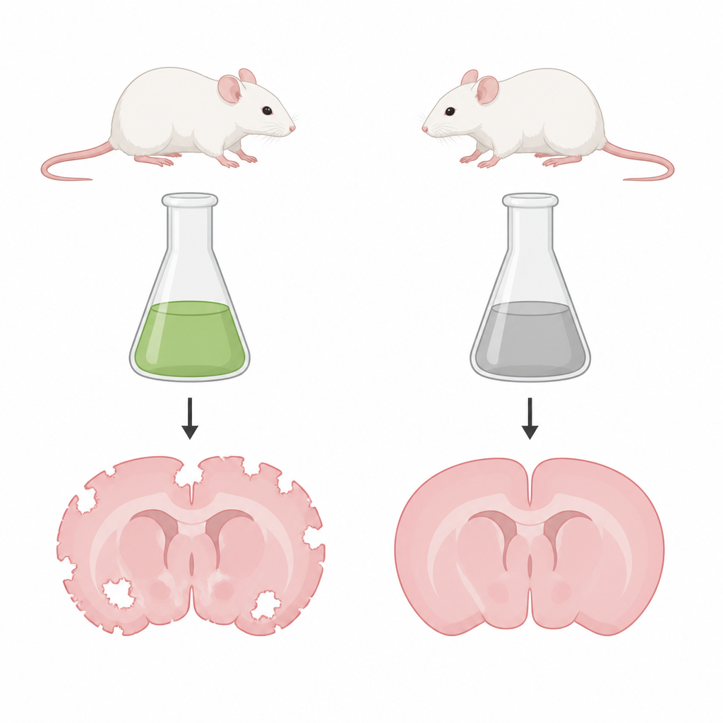
How well the brain structure was preserved
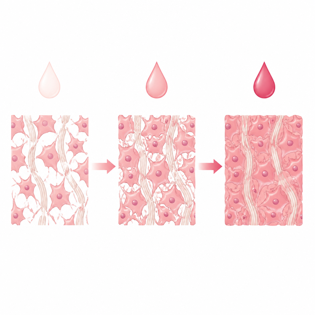
Under the microscope, formalin fixed brains showed the cleanest, most continuous tissue with only tiny cracks, clear cell outlines, and well kept layers of gray and white matter. Lactic acid samples looked worse overall, especially at low concentrations, where the tissue showed more breaks, looser nerve fibers, and blurred cell details. Stronger lactic acid solutions did noticeably better: the highest strengths reduced cracking and kept cell shapes reasonably intact, although the white matter, which is rich in fat, remained more fragile than in formalin fixed samples. When the acidity of lactic acid was neutralized, its performance dropped sharply, suggesting that its ability to preserve tissue depends in part on staying acidic.
Perfusion improves lactic acid performance
When the researchers switched from simple immersion to perfusion through the blood vessels, lactic acid’s performance improved. High strength lactic acid delivered by perfusion produced brain slices with far fewer cracks and clearer cell details than samples treated with salt solution alone, and in some respects came closer to formalin. Even so, formalin still gave the most stable white matter and the sharpest nuclear features. The patterns point to a key difference in how the chemicals work: formalin creates strong cross links between proteins, while lactic acid mainly denatures and coagulates proteins without forming the same sturdy network, leaving fat rich areas more vulnerable during later processing steps.
What this means for future practice
This study shows that lactic acid cannot fully replace formalin for brain tissue, particularly when the most delicate structures must be preserved. However, concentrated lactic acid, especially when delivered by perfusion, can keep much of the basic brain architecture intact and does so with a chemical that is far less hazardous to handle. For certain research or teaching situations where avoiding formaldehyde is a priority and some loss of fine detail is acceptable, lactic acid based fixation could offer a useful compromise between safety and tissue quality.
Citation: Venuto, M.T., Soldat-Böttcher, Z., Kleine, J. et al. Evaluation of lactic acid as a novel fixative for histological and neuroanatomical applications. Sci Rep 16, 15746 (2026). https://doi.org/10.1038/s41598-026-51513-y
Keywords: lactic acid fixation, formalin alternative, mouse brain histology, tissue preservation, neuroanatomy