Clear Sky Science · en
Identification of NF-κB pathway-related biomarkers in myocardial ischemia-reperfusion injury: based on transcriptomics analysis and RT-qPCR validation
Why saving heart muscle can still cause harm
When someone has a heart attack, doctors race to reopen the blocked artery. Ironically, restoring blood flow can itself injure the heart, a problem called ischemia‑reperfusion injury. This study asks a simple but important question: can we find reliable molecular warning lights inside heart cells that signal this damage early and point to new treatments? By combining big‑data gene analysis with animal experiments, the researchers homed in on two such signals within a major inflammation pathway.
Heart under stress from loss and return of blood
Myocardial ischemia‑reperfusion injury happens when a starved heart muscle suddenly receives blood again during procedures such as opening a clogged coronary artery or heart surgery. The rush of oxygen can trigger bursts of harmful molecules, disturb energy‑producing mitochondria, and ignite intense inflammation. White blood cells flood into the heart tissue, small blood vessels clog, and heart muscle cells die through several forms of programmed cell death. These events not only limit how much heart function is restored after treatment but also increase the risk of long‑term heart failure. Because inflammation sits at the center of this storm, pathways that control inflammatory signals are prime suspects and targets.
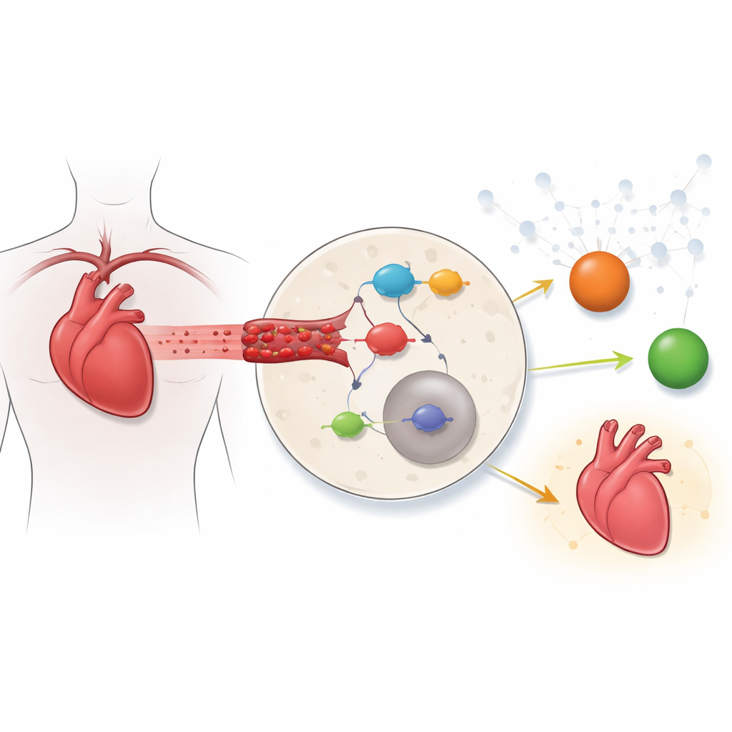
A key inflammation switch inside heart cells
One of the body’s master switches for inflammation is the NF‑κB pathway, a chain of proteins that turns on many immune and survival genes. Under calm conditions, NF‑κB is kept in check in the cell’s fluid by an inhibitor protein; under stress, that inhibitor is removed and NF‑κB moves into the nucleus to activate inflammatory programs. Earlier heart studies usually examined one or two genes or drugs at a time. Here, the authors took a more global view. They merged gene activity data from mouse hearts with a catalog of NF‑κB‑related genes to see which members of this pathway changed most strongly during ischemia‑reperfusion.
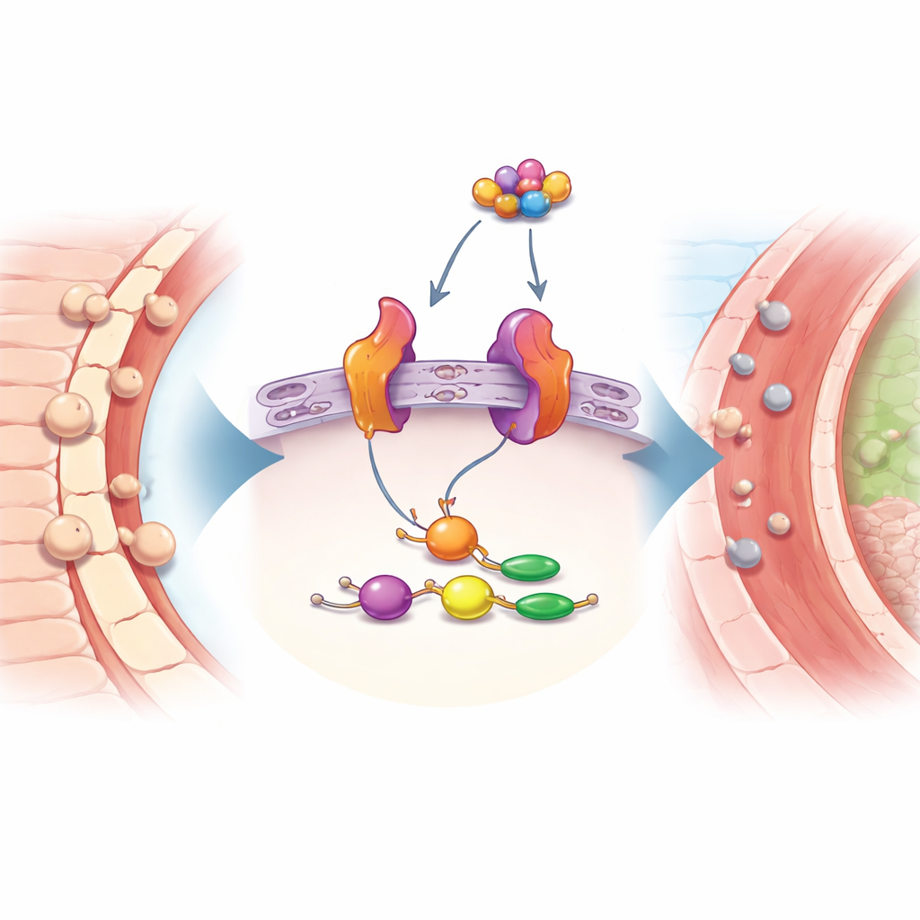
Two standout molecular warning lights
From hundreds of altered genes, only nine belonged to the NF‑κB pathway, and network analysis highlighted two of them as central players: Nfkbia and Icam1. Nfkbia encodes the key brake on NF‑κB, while Icam1 produces a sticky protein on blood vessel and immune cells that helps white blood cells attach and enter tissues. In two independent mouse datasets, both genes were consistently more active after heart injury than in control hearts. Further analysis linked Icam1 to changes in the tissue scaffold around cells, and Nfkbia to the tiny power plants inside cells, suggesting that one is tied more to the outside environment and cell‑to‑cell interactions, while the other is tied to energy balance and stress inside the cell.
Hidden RNA circuits and a possible drug candidate
Digging deeper, the team mapped how other molecules might regulate these two markers. They predicted transcription factors that switch these genes on, as well as a web of small regulatory RNAs and longer “sponge” RNAs that may fine‑tune Icam1 activity. One small RNA, called mmu‑miR‑706, stood out as a potential brake on Icam1 that could itself be blocked by several long RNAs, forming a regulatory loop that might amplify inflammation. The researchers then stepped from biology toward therapy: using drug‑gene databases and computer docking, they searched for existing compounds that might bind both NFKBIA and ICAM1. One molecule, the protease inhibitor TLCK, appeared to attach snugly to both proteins in simulations, hinting that it might calm the NF‑κB pathway and reduce white‑blood‑cell sticking in the injured heart.
Mouse experiments confirm the signal
To ensure these changes were not just artifacts of computer analysis, the team created a mouse model of ischemia‑reperfusion, briefly tying off and then reopening a heart artery. They measured gene activity in heart tissue and found that Nfkbia and Icam1 were both clearly elevated compared with uninjured control hearts. This experimental confirmation strengthens the case for these genes as robust markers of the injury process rather than random fluctuations in gene activity.
What this means for future heart care
For non‑specialists, the take‑home message is that the study identifies two molecules that act like dashboard warning lights for inflammation‑driven damage when blood flow returns to the heart. Nfkbia reflects how hard the cell is working to keep a powerful inflammation switch under control, while Icam1 reflects how strongly immune cells are being summoned into the heart tissue. Although much work remains—especially to test whether targeting these molecules or TLCK can safely protect human hearts—this research offers a clearer map of the injury process and new starting points for drugs that could make life‑saving reperfusion therapies safer and more effective.
Citation: Ting, W., Helong, X., Xiaoyu, W. et al. Identification of NF-κB pathway-related biomarkers in myocardial ischemia-reperfusion injury: based on transcriptomics analysis and RT-qPCR validation. Sci Rep 16, 11729 (2026). https://doi.org/10.1038/s41598-026-47878-9
Keywords: heart reperfusion injury, NF-kappaB inflammation, cardiac biomarkers, gene expression analysis, ICAM1 and NFKBIA