Clear Sky Science · en
The study of nomogram model based on CT radiomics and clinical features for histological classification of parotid gland tumors
Why this matters for patients and doctors
When a lump appears near the jaw, both patients and doctors urgently want to know: Is it harmless or dangerous, and what treatment is really needed? For tumors of the parotid gland, the largest salivary gland, the answer usually comes only after surgery or an invasive needle test. This study explores whether information already hidden in routine CT scans, combined with simple patient details, can sort these tumors into key types in advance, helping tailor surgery and avoid unnecessary risks.
The problem of look‑alike gland tumors
Parotid gland tumors are relatively rare but clinically important. Most are benign, especially three common types: pleomorphic adenoma, Warthin tumor, and basal cell adenoma. A smaller share are malignant, requiring more aggressive surgery and sometimes radiation. Unfortunately, many of these tumors look similar on standard imaging and often have no distinctive symptoms. Doctors rely on fine‑needle aspiration biopsies and their own reading of CT or MRI scans, but biopsies can be uncomfortable, sometimes inconclusive, and carry small risks. This creates a strong motivation for better, non‑invasive tools that can distinguish tumor types before the surgeon ever picks up a scalpel.
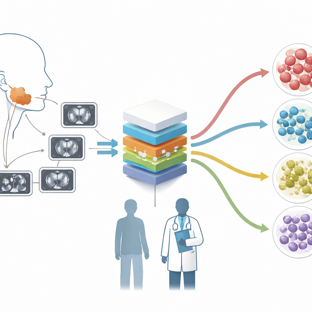
Turning CT scans into rich data maps
The researchers used a technique called radiomics, which treats each CT scan as a rich data source rather than just a picture. From enhanced CT images of 427 patients at two hospitals, they painstakingly outlined each tumor and fed the images into specialized software that extracts over a thousand numerical features. These features describe patterns of brightness, texture, and shape that the human eye cannot easily quantify. To make the data reliable across different scanners, the team resampled and normalized the images and used a statistical method called ComBat to harmonize features from multiple machines.
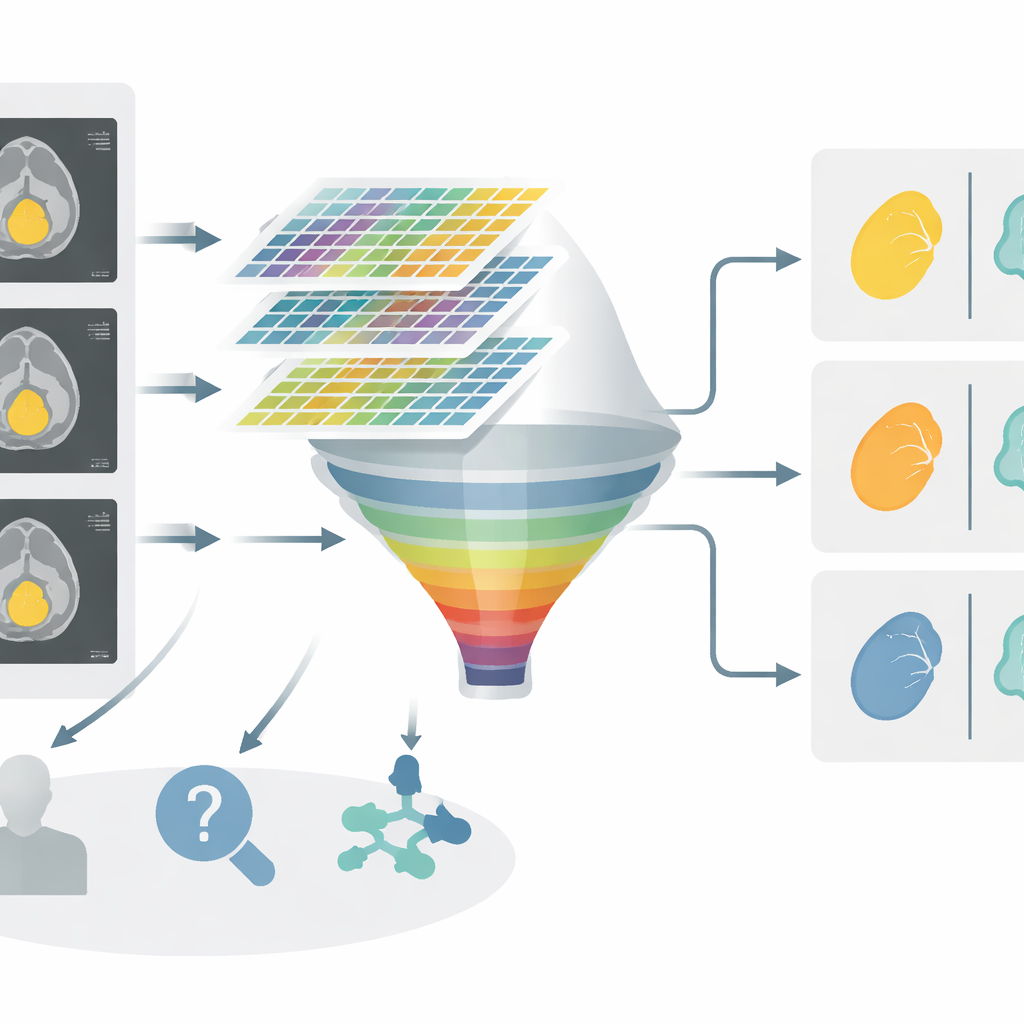
Building smart models in three decision steps
Instead of trying to classify every tumor type at once, the team broke the problem into three simple decisions that mirror how a clinician might think. First, they asked: benign or malignant? Second, among benign tumors, is this pleomorphic adenoma versus the other two types combined? Third, if it is one of those two, is it Warthin tumor or basal cell adenoma? For each step, they used machine‑learning methods—logistic regression, support vector machines, and gradient‑boosted trees—to find the most informative radiomics features and build a prediction model. They evaluated these models by how well they separated the groups, measuring performance with the area under the ROC curve, a standard score for diagnostic tests.
Blending image patterns with everyday clinical clues
While radiomics captured subtle image details, the authors also recognized the value of basic clinical information. They assessed features such as symptoms, tumor margin, lymph node enlargement, age, tumor size, and cyst‑like areas on CT. Using statistical analysis, they identified which of these factors truly contributed to distinguishing the tumor groups. Then they combined these clinical predictors with the radiomics "signature" into a single graphical tool called a nomogram. This nomogram turns complex model output into an easy‑to‑use point system that can estimate the probability of each tumor type for an individual patient.
What the new tool achieved
Across all three decision steps, the combined nomogram outperformed models that used only clinical features or only radiomics. For example, when separating benign from malignant tumors, the integrated model showed better overall accuracy and a higher diagnostic score than clinical factors alone. Similar improvements appeared in the two benign‑to‑benign comparisons, where subtle differences are especially hard to see by eye. Decision‑curve analysis, which weighs the benefits of correct classification against the harms of being wrong, suggested that the nomogram could offer more net clinical benefit across a wide range of real‑world decision thresholds.
What this means going forward
In plain terms, the study shows that careful analysis of CT images, paired with routinely collected patient details, can help doctors more confidently sort parotid gland tumors into meaningful categories before surgery. The tool does not replace biopsy or expert judgment, but it could guide decisions about how extensive an operation should be and which patients might safely avoid overly aggressive treatment. With larger, multi‑center studies and the inclusion of other imaging types, such radiomics‑based nomograms may become a practical part of everyday care, bringing more personalized and less invasive diagnosis to people with salivary gland tumors.
Citation: Shen, Q., Liu, Y., Xu, F. et al. The study of nomogram model based on CT radiomics and clinical features for histological classification of parotid gland tumors. Sci Rep 16, 11665 (2026). https://doi.org/10.1038/s41598-026-46970-4
Keywords: parotid gland tumors, CT radiomics, machine learning diagnosis, tumor classification, noninvasive imaging