Clear Sky Science · en
Feasibility of 68Ga-FAPI-46 PET for evaluating active fibrosis in aortic aneurysm
Why weak spots in a major artery matter
Aortic aneurysms are silent weak spots in the body’s main artery that can suddenly tear or burst, often without warning. Doctors currently judge danger mostly by measuring how wide the artery has stretched over time. But size alone does not reveal how "active" or fragile the vessel wall really is. This study explores a new type of medical scan that might show when the aneurysm tissue is biologically active and potentially at higher risk, long before a disaster occurs. 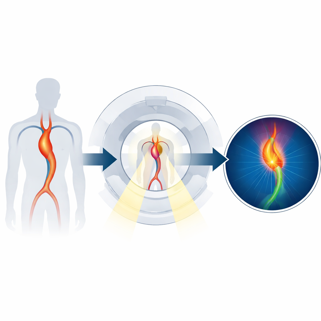
The hidden players in aortic damage
Inside the wall of the aorta, fibroblasts act like maintenance workers, building and repairing the supporting scaffold around blood vessels. When injury or inflammation lingers, these cells can switch into an overdrive state, helping to lay down stiff scar-like tissue called fibrosis. Over time, this remodeling process can weaken the vessel wall, contributing to the growth of an aneurysm. The researchers focused on a surface molecule called fibroblast activation protein, or FAP, which appears when fibroblasts are switched on and busily reshaping tissue.
A tracer that lights up active scarring
To see FAP inside the body, scientists have developed small molecules known as FAPI tracers that seek out and bind to FAP. In this study, the team used a version labeled with a radioactive element, gallium-68, and imaged it with positron emission tomography (PET). When injected into the bloodstream, this tracer should collect where activated fibroblasts are most abundant, causing those regions to glow on the scan. The central question was whether this glow in the aortic wall would match actual signs of fibrosis and whether it would be stronger in more aggressive aneurysms. 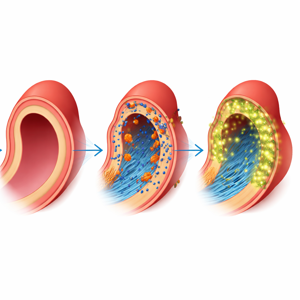
What the tissue and scans revealed
The investigators prospectively enrolled 20 patients who were already scheduled for surgery to remove sections of their diseased aorta, and they compared them with 9 people scanned for lung cancer who had no known vessel disease. All aneurysm patients received a gallium-68 FAPI-46 PET/CT scan before surgery. In the operating room, surgeons collected samples from the aneurysm wall and, when possible, from more distant, apparently normal parts of the aorta. These samples were carefully processed to measure FAP and other molecules, to visualize fibrosis under the microscope, and to test how much tracer the tissue could bind. The researchers also reviewed past CT scans to calculate how fast each aneurysm had grown over time.
Stronger signals in more active aneurysms
The surgical specimens showed that aneurysm tissue contained much more FAP and another remodeling signal, transforming growth factor beta, than the remote, non-aneurysm aorta. Fibrous collagen strands appeared not only on the outer layer but also deeper into the wall, consistent with advanced scarring. Importantly, the intensity of the PET signal—the maximum tracer uptake in the aneurysm—tracked with how much FAP the tissue actually expressed. Patients with aneurysms had higher PET uptake in their aorta than the control group, whose vessels showed only low background signal. Within the aneurysm group, higher visual grades and higher uptake values were associated with faster annual growth of the aneurysm, even though most patients overall had relatively slow enlargement.
What this could mean for patient care
Taken together, the results suggest that gallium-68 FAPI-46 PET can noninvasively highlight areas of active fibrosis in the aortic wall and that this activity relates, at least modestly, to how quickly an aneurysm is growing. Instead of relying only on size and repeated CT measurements, doctors might one day use this kind of scan to gauge how biologically "hot" an aneurysm is and to refine decisions about when to intervene. The authors caution that their study was small and included a mix of aneurysm locations, so larger, more focused trials are needed. Still, the work points toward a future in which the risk of aortic rupture could be judged not just by how big the vessel looks, but by how intensely its hidden repair machinery is working.
Citation: Suh, H.Y., Byun, J.W., Lee, SP. et al. Feasibility of 68Ga-FAPI-46 PET for evaluating active fibrosis in aortic aneurysm. Sci Rep 16, 14115 (2026). https://doi.org/10.1038/s41598-026-44481-w
Keywords: aortic aneurysm, fibrosis imaging, PET scan, fibroblast activation protein, vascular disease