Clear Sky Science · en
Deep learning for predicting stem cell efficiency for use in beta cell differentiation
Why this research matters for future diabetes care
For people with type 1 diabetes, an emerging dream is to replace lost insulin-producing cells instead of relying on lifelong injections. Scientists can now grow such cells from a patient’s own stem cells, but the process is slow, expensive and often fails. This study shows how artificial intelligence can look at early microscope images of stem cells and predict, within just a couple of days, which batches are likely to become good insulin-producing cells and which are doomed to disappoint—potentially saving time, money and precious biological material.
From skin cells to insulin-making cells
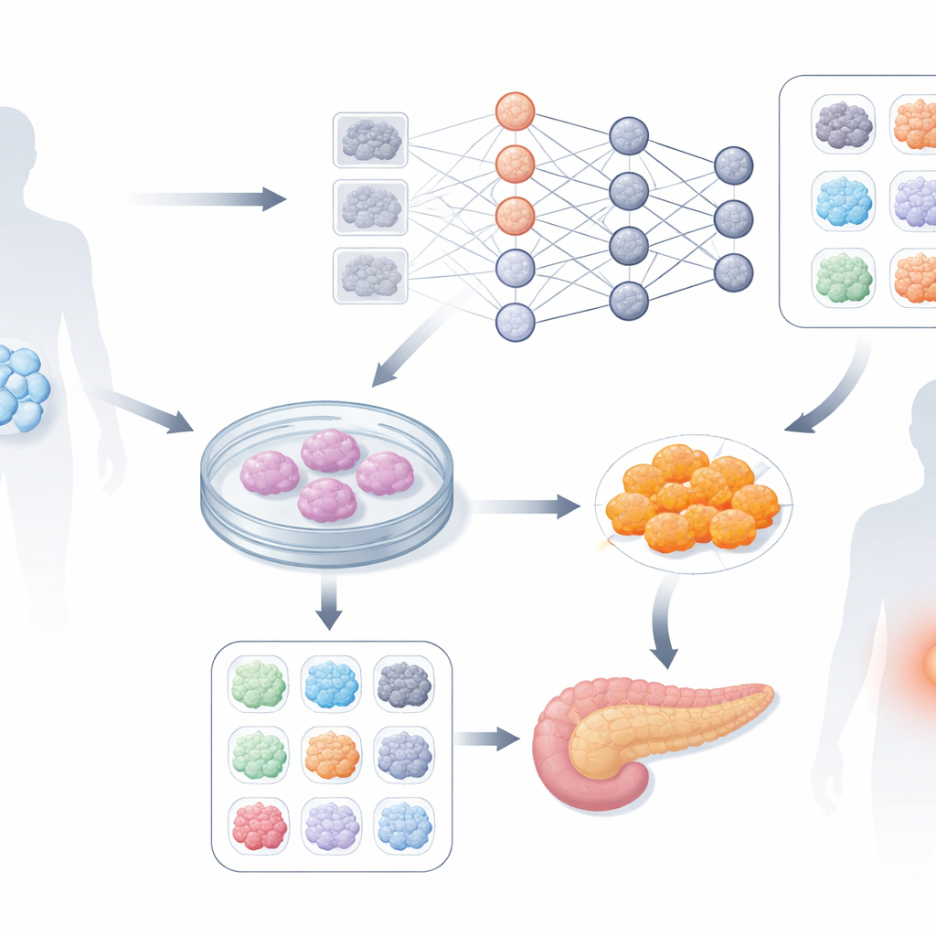
In modern cell therapy research, ordinary adult cells from a patient can be reprogrammed back into a stem-cell-like state, called induced pluripotent stem cells. These stem cells can then be guided through a month-long process to become pancreatic beta cells, the cells that release insulin. In theory, this provides a personal, rejection-proof source of replacement cells for people with type 1 diabetes. In practice, each patient’s stem cell “clone” behaves differently: some yield many healthy beta cells, others very few. Because the differences are subtle at the beginning, even skilled experts cannot reliably tell early on which clones will perform well. That uncertainty forces researchers to carry weak clones all the way through the 34-day procedure, wasting effort and raising costs.
Teaching computers to read subtle cell patterns
The authors set out to see whether a deep learning system—similar in spirit to those used in image recognition—could spot early visual cues in simple, label-free microscope images. They worked with six patient-derived stem cell clones, all headed toward the same target cell type. Instead of using colored stains or complex imaging, they relied on standard phase-contrast pictures taken every hour during the first four days of development. Later, the success of each clone was measured using a known marker on day four that correlates with how well the cells eventually turn into functioning beta cells. Clones were grouped into “good” and “bad” based on this marker, and the neural network was trained to tell these groups apart from early images alone.
How well the system can foresee success
Using an EfficientNet-based deep learning model, the researchers chopped high-resolution images into smaller patches and trained the network on many such pieces from each clone. They evaluated performance in two ways: how often individual patches were correctly classified, and how often the overall prediction for a whole clone was right, using a voting scheme. While images taken before differentiation began contained no useful visual differences, by around two days into the process the network’s forecasts became strikingly accurate. At around 53 hours, the system correctly identified the eventual outcome for 96.7 percent of clones, providing a strong early warning signal about which stem cell batches are worth continuing and which could be halted and restarted with new cells.
What the computer “looks” at in the cells
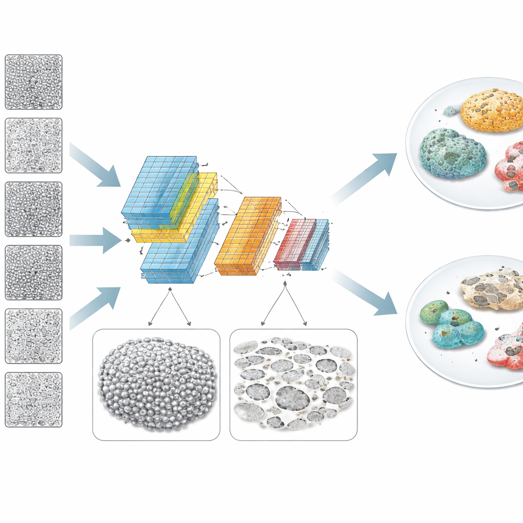
To move beyond a black-box prediction, the team used explainable-AI tools and traditional image analysis to understand what visual features mattered. Heatmaps of the network’s attention suggested that it was not focusing on individual cells, but on broader patterns across the cell sheet. Good clones tended to form more continuous, uniform layers of cells, while bad clones showed more empty patches or “holes” and uneven brightness across regions spanning tens to hundreds of micrometers. A separate mathematical analysis of image structure backed this up: for bad clones, intensity variations were stronger over intermediate length scales, consistent with patchy coverage and debris. The model’s explanations correlated with simple measures such as brightness variance, and the authors showed how different image normalization strategies affected performance depending on how clean or debris-filled the culture was.
Steps toward more efficient and affordable cell therapy
This work is an early but promising demonstration that deep learning, combined with very simple live-cell imaging, can act as an automated early-quality inspector for stem cell production. By flagging weak clones after only a couple of days, such systems could cut down on wasted culture runs and lower the cost of producing patient-specific beta cells, helping bring cell therapy for type 1 diabetes closer to routine clinical use. Although the study involved only six clones and will need to be validated on larger and more diverse datasets, it shows that subtle, population-level cell patterns contain rich information that computers can exploit long before the human eye can see clear differences.
Citation: Schöb, F.J., Binder, A., Zamarian, V. et al. Deep learning for predicting stem cell efficiency for use in beta cell differentiation. Sci Rep 16, 12788 (2026). https://doi.org/10.1038/s41598-026-42830-3
Keywords: type 1 diabetes cell therapy, induced pluripotent stem cells, deep learning microscopy, beta cell differentiation, image-based quality control