Clear Sky Science · en
Localization of Gli1-expressing cells after pulp revascularization and their involvement in newly formed mineralized tissue in mice
Why Saving Young Teeth Matters
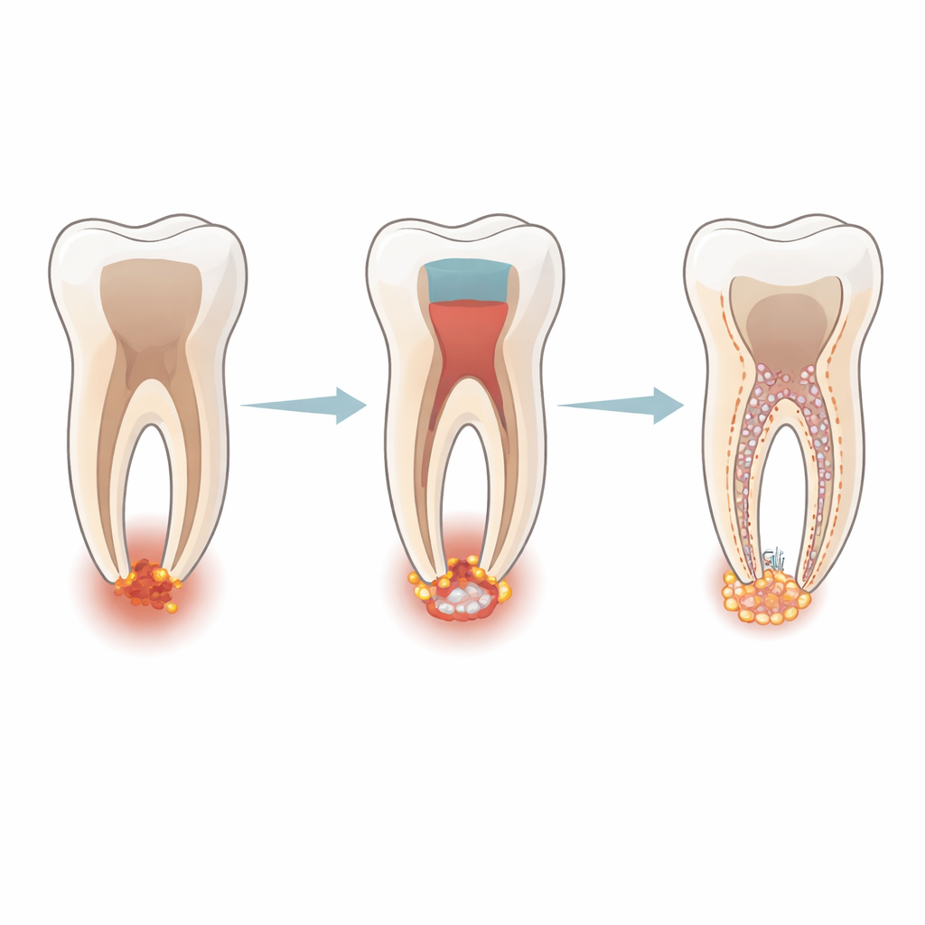
When the soft core of a young tooth dies after deep decay or an accident, dentists face a dilemma: traditional treatments can close the root tip but often leave the tooth fragile and prone to breaking. Newer "regenerative" procedures aim not just to seal the tooth, but to coax it to keep growing and strengthen itself from within. This study in mice asks a key question for making those treatments reliable: which cells actually move into the emptied root canal and lay down the new hard tissue that stabilizes the tooth?
A New Look at Tooth Healing
The researchers focused on a special group of repair cells marked by a protein called Gli1. These cells normally sit around the tip of the tooth root in the tissues that anchor the tooth to the jaw. Using genetically modified mice in which Gli1-expressing cells glow under the microscope, the team followed where these cells travel after a pulp revascularization–style procedure that mimics regenerative treatment in the clinic. In this procedure, the pulp of an immature molar is removed, the root canal is cleaned, and a blood clot is deliberately triggered at the open root tip before the tooth is sealed.
Watching Repair Cells on the Move
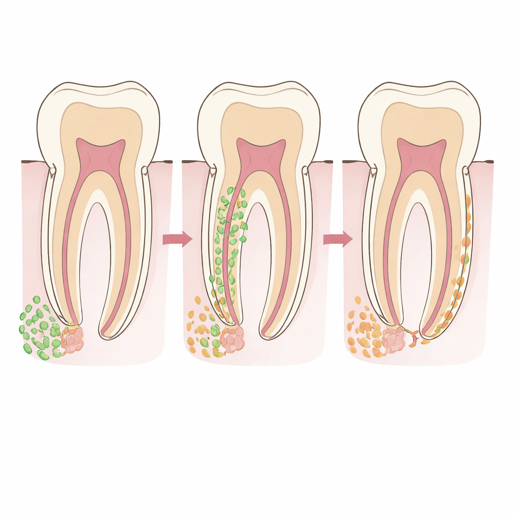
Over three weeks, the scientists examined tooth sections at several time points. Right after treatment, the emptied root canal contained almost no Gli1-marked cells; instead, these cells clustered in the tissue just beyond the root tip. By three days, the canal—especially its lower part—was packed with cells, and Gli1-marked cells formed a continuous bridge from the region around the root tip into the canal. Over the next days, these cells gradually advanced toward the upper canal, lining the canal walls and eventually appearing all the way up to the area just beneath the sealing material placed by the dentist.
From Traveling Cells to New Hard Tissue
The team then asked whether these migrating cells were actually responsible for building new mineralized tissue. They looked for another marker, osterix, which is typical of cells that form cementum, the hard layer that coats the root surface. Many of the Gli1-marked cells also displayed this cementum-related marker, first near the root tip and later inside the canal, suggesting they were switching from a mobile repair state into active tissue builders. As time went on, small islands and bands of new hard material appeared along the canal walls, continuous with the tooth’s original root coating. Gli1-marked cells, including some that no longer showed the early cementum marker, were found around and within this new tissue, consistent with them having produced it and then become embedded as long-term resident cells.
Ruling Out Other Tooth-Building Paths
To pinpoint the source of the new material, the authors checked several alternative cell types. A marker typical of the periodontal ligament’s fibrous support cells, periostin, was largely absent from the areas where new tissue formed, suggesting that the usual ligament cells along the side of the root were not the main contributors. Likewise, a marker associated with dentin-making cells inside the pulp, nestin, did not appear in the treated canals, even though it was clearly visible in untouched neighboring roots. Combined with the close continuity between the new material and the existing cementum, these findings argue that the fresh mineralized layer inside the canal is cementum-like rather than dentin-like, and that it is built predominantly by Gli1-marked cells that arrive from the root tip region.
What This Means for Future Dental Care
For patients, the key message is that successful regenerative root treatments may depend heavily on mobilizing the right kind of local repair cells at the root tip, rather than reviving classic pulp cells deep inside the tooth. In this mouse model, Gli1-expressing cells around the root tip multiply briefly, move into the cleaned canal, adopt a cementum-forming role, and lay down a new inner shell of hard tissue that thickens and lengthens the root. Understanding and eventually guiding this process could help dentists design regenerative procedures that not only keep infected young teeth in the mouth, but also rebuild them into stronger, more fracture-resistant structures.
Citation: Tashiro, K., Ikarashi, T., Haketa, M. et al. Localization of Gli1-expressing cells after pulp revascularization and their involvement in newly formed mineralized tissue in mice. Sci Rep 16, 10065 (2026). https://doi.org/10.1038/s41598-026-40945-1
Keywords: pulp revascularization, tooth regeneration, periodontal ligament cells, cementum formation, dental stem cells