Clear Sky Science · en
Bevacizumab-IRDye800 as an imaging probe for the detection of prostate cancer in mice
Seeing Hidden Tumors
Prostate cancer is common, yet spotting small or early tumors inside the body is still difficult. Doctors rely on blood tests, scans, and tissue samples, but each has limits. This study explores a new way to “light up” prostate tumors in living animals using a glowing drug, with the long term goal of helping surgeons and radiologists see cancer more clearly and remove it more precisely.
A Glow-in-the-Dark Target
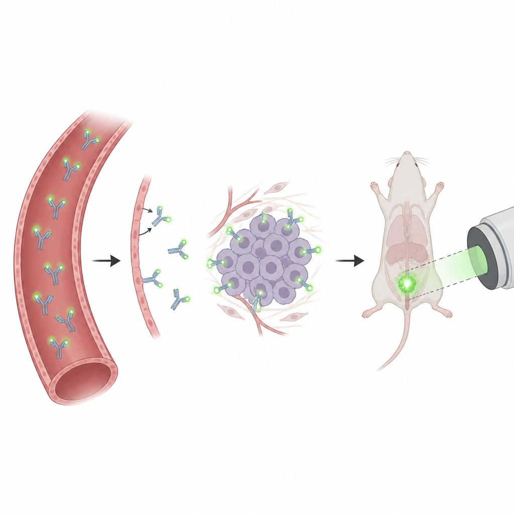
The researchers focused on a drug called Bevacizumab, already used in patients to block the growth of blood vessels that feed tumors. Many prostate cancers produce high levels of a signal protein that Bevacizumab recognizes. By attaching a near-infrared fluorescent dye to this antibody, the team created a probe that should travel through the bloodstream, latch onto prostate cancer cells and nearby new blood vessels, and give off light that special cameras can detect through the body.
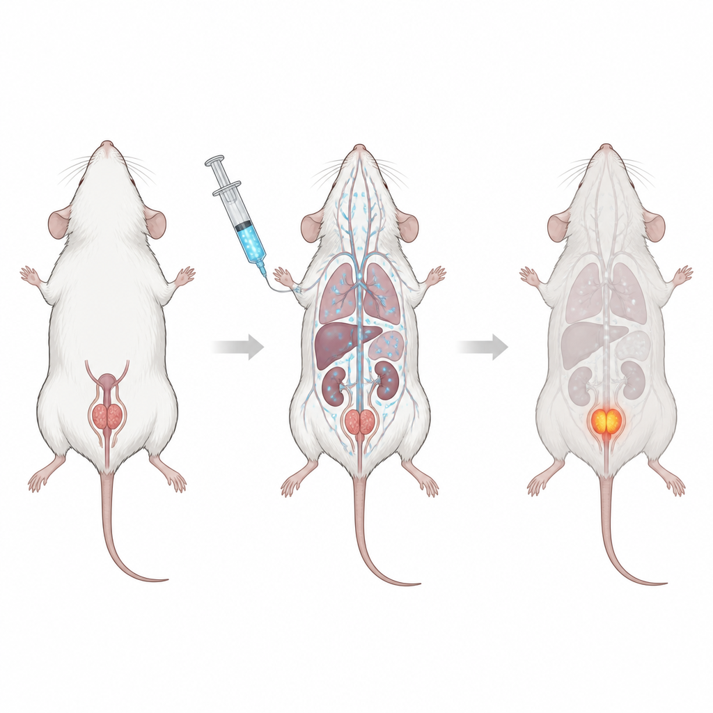
Testing in a Mouse Stand-In for Human Disease
To test this idea, the team built a realistic mouse model of human prostate cancer. Human prostate cancer cells were engineered to glow by bioluminescence so that their growth could be tracked independently. These cells were then injected directly into the mouse prostate, where they reliably formed local tumors. After tumors had developed, the mice received the fluorescent Bevacizumab probe through a vein. The scientists then used different imaging systems to follow where the probe went over time and to find the best moment when tumor signal was high and background signal was low.
Lighting Up Primary Tumors and Spread
The researchers compared their targeted probe with indocyanine green, a common non-specific fluorescent dye already used in surgery. While both groups of mice had similar tumor sizes, only the Bevacizumab probe produced a strong fluorescent signal at the prostate location 72 hours after injection. Three-dimensional imaging showed clear accumulation of the probe in the prostate, and when the prostates were removed, the fluorescent signal closely overlapped with the independent bioluminescent signal from the cancer cells. Under the microscope, the antibody probe could be seen within the tumor tissue, especially around blood vessels. In a second model, where cancer cells were allowed to spread through the bloodstream, the probe also reached some metastatic sites such as the liver, again matching areas where tumor cells were present.
What This Could Mean for Patients
Current advanced imaging for prostate cancer often relies on radioactive tracers and costly scanners, which are not ideal for guiding routine biopsies or surgery. Because Bevacizumab is already approved for human use and has been tested as a fluorescent probe in other cancers, this work suggests it could be adapted to help doctors see prostate tumors and some metastases with near-infrared cameras. In the mouse models, the fluorescent Bevacizumab probe attached specifically and in high amounts to tumor areas, unlike a non-targeted dye. While more studies and human trials are required, the findings indicate that a glowing version of an existing drug may one day help clinicians find and remove prostate cancer more accurately, improving diagnosis and treatment without adding radiation exposure.
Citation: Genevois, C., Dugot-Senant, N., Canron, MH. et al. Bevacizumab-IRDye800 as an imaging probe for the detection of prostate cancer in mice. Sci Rep 16, 15108 (2026). https://doi.org/10.1038/s41598-026-39705-y
Keywords: prostate cancer imaging, fluorescent antibody, bevacizumab, near infrared, mouse model