Clear Sky Science · en
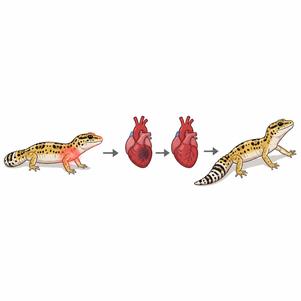
Heart ventricle regeneration in the lizard Eublepharis macularius, the leopard gecko
How a small lizard points to self-healing hearts
Heart attacks in people often leave behind permanent scar tissue that weakens the heart. Scientists are hunting for animals that can instead rebuild healthy heart muscle, hoping to uncover clues that might one day guide new treatments. This study turns to an unexpected ally: the leopard gecko, a popular pet lizard that is already famous for regrowing its tail, skin, and even parts of its brain.
A lizard with more than a regrowing tail
Leopard geckos are standout regenerators among backboned animals that lay eggs on land, a group that includes reptiles, birds, and mammals. Beyond their detachable tails, they can repair spinal cord, skin, teeth, and brain tissue. Yet almost nothing was known about how their hearts respond to serious injury. The researchers set out to learn whether a gecko’s heart simply scars, like a typical mammal, or can truly rebuild lost muscle, more like certain fish and salamanders that are known to regenerate their hearts.
Freezing part of the heart to test repair
To mimic a heart attack, the team gently opened the chest of anesthetized geckos and briefly touched a liquid nitrogen–chilled metal probe to the pumping chamber of the heart. This froze and killed about one fifth of the ventricle, creating a sharply defined patch of dead tissue similar in effect to a severe blockage in a coronary artery. Every animal survived the operation, allowing the scientists to track what happened over days and months using tissue stains, cell markers, and ultrasound scans that measure how well the heart squeezes.
From cell death to new muscle
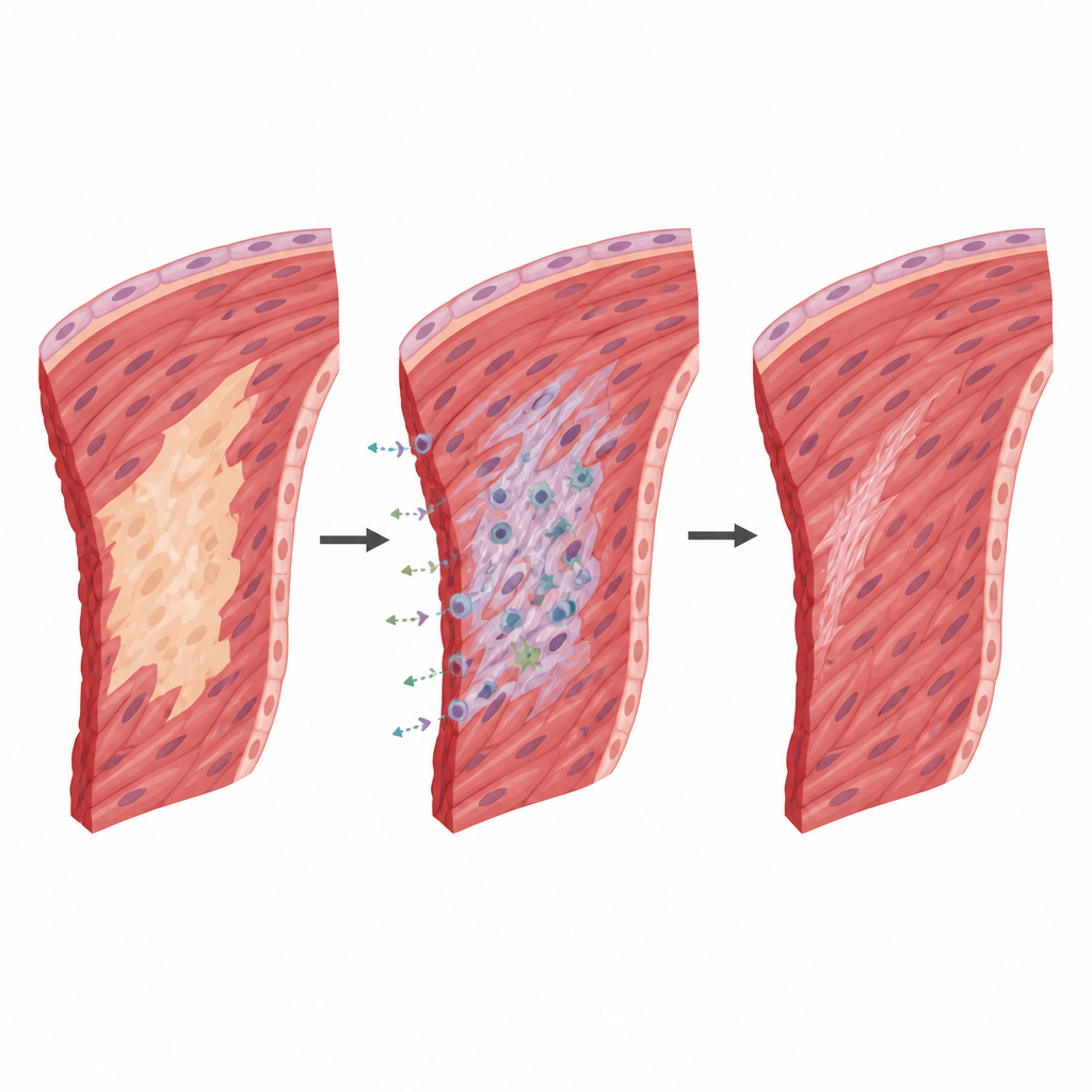
In the first days after injury, the damaged region was filled with dead cells and invaded by immune and support cells, along with early collagen fibers that form a temporary internal “patch.” At this stage, heart muscle cells were missing from the core of the injury. Soon after, however, the researchers saw a surge of cell division in surviving heart muscle cells around the wound, as well as in nearby non-muscle cells. This burst of growth was strongest next to the damaged zone and faded with distance, suggesting the heart recruits local cells to rebuild the injured wall. Over several weeks, the collagen mesh became more organized and heart muscle gradually grew back into the damaged area.
Heart function rebounds as the scar shrinks
Immediately after the freeze injury, the hearts pumped less efficiently. Ultrasound images showed that the ventricle’s squeezing power dropped and remained low for several weeks. By about 100 days, though, pumping strength had returned to the same level as in uninjured and sham-operated geckos. Detailed pressure measurements inside the ventricle confirmed that both the contraction and relaxation phases of each heartbeat were restored. At this late stage, only a tiny sliver of fibrous tissue remained at the edge of the heart wall, covering less than 2 percent of its cross section, while the rest of the damaged region had been replaced by new muscle.
Shared molecular playbook across distant species
To see which genes switch on during this repair, the team analyzed RNA from tissue bordering the injury at several time points. They found thousands of genes whose activity rose or fell compared with sham surgery. Many of these are known from fish and salamander studies to take part in heart development, cell division, energy metabolism, and the building and reshaping of the supporting matrix between cells. Early on, genes tied to wound healing and collagen production were boosted, later giving way to those involved in forming new muscle and blood vessels. This pattern suggests that, despite hundreds of millions of years of evolution, the leopard gecko taps into a molecular toolkit that resembles that of other animals capable of rebuilding heart tissue.
What this means for future heart repair
The study shows that an adult leopard gecko can regrow most of the damaged pumping chamber of its heart and regain near-normal function after a severe injury. While this does not translate directly into human treatment, it broadens the roster of species that can naturally replace lost heart muscle. By comparing the steps and genetic signals used by geckos, fish, salamanders, and mammals, researchers can better understand which features of heart regeneration are widely shared and which are unique. That knowledge could eventually guide strategies to encourage more robust repair in the human heart after injury.
Citation: Jacyniak, K., Williams, C.J.A., Beaufrère, H. et al. Heart ventricle regeneration in the lizard Eublepharis macularius, the leopard gecko. npj Regen Med 11, 22 (2026). https://doi.org/10.1038/s41536-026-00469-8
Keywords: heart regeneration, leopard gecko, cardiac repair, regenerative biology, myocardial injury