Clear Sky Science · en
Imaging and genome-supported association of glymphatic system function and multiregional brain characteristics with Parkinson’s disease
Why this brain-cleansing study matters
Parkinson’s disease is best known for tremors and stiffness, but long before these symptoms appear, subtle changes are already unfolding deep in the brain. This study explores a hidden “cleaning service” in the brain that helps wash away waste, and shows how its breakdown—together with tiny changes in brain tissue—may signal Parkinson’s disease years earlier than current tests can. Understanding and measuring these changes noninvasively could open the door to earlier diagnosis and more precisely targeted treatment.
The brain’s night-shift cleaning crew
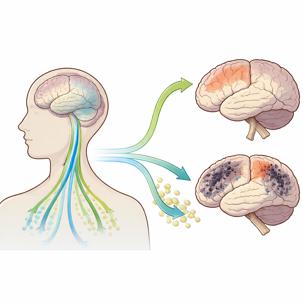
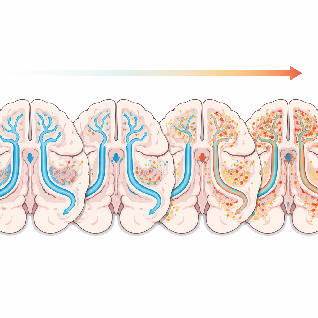
Our brains constantly produce waste products that need to be cleared away, including proteins like alpha-synuclein, which can clump and damage nerve cells in Parkinson’s disease. A recently described network, called the glymphatic system, circulates fluid along blood vessels to rinse out this waste. The authors used a diffusion MRI measure known as the ALPS index to estimate how well this cleaning system is working. They also used another MRI measure, called free water, which captures how much freely moving fluid is present in different brain regions—a sign of microscopic damage, swelling, or inflammation. By combining these two views of the brain, the team asked: can we better detect early Parkinson’s disease, and can genetics tell us which brain regions truly contribute to risk?
What the brain scans revealed
The researchers analyzed data from 118 people with early Parkinson’s disease and 58 healthy volunteers in a large international study. People with Parkinson’s showed a lower ALPS index, meaning poorer apparent glymphatic function, and higher free water in several brain regions. The strongest free-water changes appeared in the temporal lobe, with notable increases also in the frontal, parietal, occipital lobes and cerebellum, while deeper structures such as the caudate and thalamus did not differ much. A lower ALPS index was linked to worse movement scores, hinting that reduced brain cleansing goes hand in hand with more severe motor problems, even at an early stage.
Building a better early-warning model
Next, the team tested how well these MRI measures could distinguish Parkinson’s patients from healthy controls. On their own, both the ALPS index and temporal lobe free water showed moderate predictive power. When added to basic information such as age, sex, and education, each improved the accuracy of a simple clinical model. But the real gain came from combining the ALPS index with temporal lobe free water: together they produced the most accurate risk estimate, outperforming either measure alone. Using this pair of imaging markers, the authors built a visual scoring tool, called a nomogram, that translates a person’s scan values into an individualized probability of having Parkinson’s disease, with good internal consistency on statistical checks.
What genetics say about vulnerable brain regions
To move beyond simple association, the researchers turned to a genetic strategy called Mendelian randomization, which uses naturally occurring genetic differences as a kind of “built-in experiment.” Drawing on large genome-wide datasets, they asked whether inherited traits linked to specific brain structures actually help cause Parkinson’s disease. They found that several structural features, especially in the frontal and temporal lobes, showed a positive causal relationship with Parkinson’s risk. In other words, genetic variants that nudge these regions’ structure in particular directions were also linked to higher odds of developing the disease. Surprisingly, the ALPS index itself did not show a clear genetic causal effect on Parkinson’s onset, suggesting that reduced glymphatic function may be more important for how fast the disease progresses than for triggering it in the first place.
What this means for future care
Taken together, the findings paint a picture in which a faltering brain-cleaning system and early microstructural changes—especially in the frontal and temporal lobes—travel together with Parkinson’s disease. While the combined imaging model is not accurate enough to serve as a stand-alone diagnostic test, it could become a valuable add-on when symptoms are vague and doctors need extra evidence. The work also highlights the frontotemporal regions as promising targets for future therapies aimed at slowing cognitive and emotional problems in Parkinson’s disease. With larger and longer-term studies, standardized scan methods, and refined imaging tools, this brain-cleansing perspective may help shift Parkinson’s care toward earlier detection and more personalized intervention.
Citation: Ye, Z., Lin, Y., Lu, Y. et al. Imaging and genome-supported association of glymphatic system function and multiregional brain characteristics with Parkinson’s disease. npj Parkinsons Dis. 12, 103 (2026). https://doi.org/10.1038/s41531-026-01314-x
Keywords: Parkinson’s disease, brain imaging, glymphatic system, temporal lobe, early diagnosis